Eye fundus image quality evaluation method based on human vision system

A technology of human visual system and fundus image, which is applied in the field of medical image processing, can solve the problems of not using human visual system, improving image quality classification performance, and not being able to apply data sets, so as to avoid subjective factors and reduce data resources and time consumption, the effect of good generalization ability

Pending Publication Date: 2019-08-02
南京星程智能科技有限公司
View PDF4 Cites 18 Cited by
  • Summary
  • Abstract
  • Description
  • Claims
  • Application Information

AI Technical Summary

Problems solved by technology

For example, image quality classification using handcrafted features including shape, color, brightness, and prior knowledge, which are based on general or structural quality parameters, have poor generalization ability on new datasets and cannot be applied to larger datasets.
On the other hand, although experts rely on the human visual system's ability to identify poor-quality fundus images and can be adapted to new datasets, such assessments are highly subjective in practice.
In addition, current handcrafted feature-based methods cannot exploit the characteristics of the human visual system to improve the performance of image quality classification.

Method used


Image

  • Eye fundus image quality evaluation method based on human vision system
  • Eye fundus image quality evaluation method based on human vision system
  • Eye fundus image quality evaluation method based on human vision system

Examples


Embodiment 1

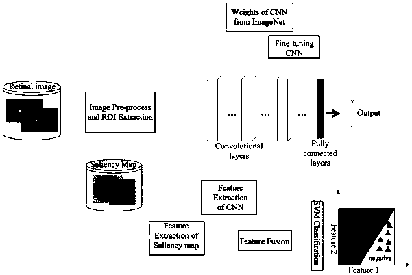
[0030] Embodiment 1: The fundus image quality assessment method based on the human visual system provided by the present invention classifies the quality of fundus images, and the specific operations are performed as follows:

[0031] 1. Select the data set;

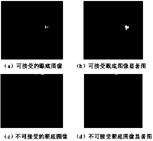
[0032] The Kaggle data set contains 80,000 diabetic retinopathy images. The image quality labels are marked by professionals into two categories: 0 means unacceptable quality fundus images, and 1 means fundus images of acceptable quality. Because the proportion of unacceptable images in all images is very small, 3864 original samples are randomly selected from the data set as the training set, and 1200 original samples are randomly selected as the test set. The training set contains 2092 samples with label 1 and 1772 samples with label 0, and the test set contains 582 samples with label 1 and 618 samples with label 0.

[0033] 2. Data preprocessing

[0034] The maximum between-cluster variance method is used to select the optim...


PUM


Abstract

The invention discloses an eye fundus image quality evaluation method based on a human vision system, which comprises the following steps: selecting a part of eye fundus images in a data set as original data samples, preprocessing the original data samples, removing a background part, and extracting an interested region part; calculating a saliency map of the fundus image; training the convolutional neural network by using the fine-tuning deep neural network, and migrating network parameters of the natural image to the training of the medical image network; extracting features of the fundus image and saliency features in the saliency map, and carrying out feature fusion; and constructing a feature matrix of the sample for the fused features, training a support vector machine classifier byutilizing the feature matrix, and classifying the quality of the fundus image. Supervision information based on a convolutional neural network and non-supervision information based on a saliency map are fused, a fusion information training classifier is utilized to classify the image quality, and a transfer learning principle is utilized to improve the performance of the image quality classification by using a method of finely adjusting the deep convolutional neural network.

Description

Technical field [0001] The present invention relates to the technical field of medical image processing, in particular to fundus image quality assessment based on the human visual system. Background technique [0002] Fundus image quality evaluation is a fundamental issue in the development of clinical assisted diagnosis of fundus images. In an automated eye disease lesion screening system, the quality of input images plays a vital role in the results of automatic screening and diagnosis of eye diseases. The automatic diagnosis and analysis system requires that the input retinal fundus images have the lowest quality that can meet the analysis requirements, so that it is convenient to extract features for subsequent diagnosis requirements. However, in practice, due to factors such as the professional knowledge level of the operators, the different types of equipment used, and the patient's condition, the acquired fundus images will have image quality problems such as noise, blur, ...

Claims


Application Information

Patent Timeline
Patent Type & Authority Applications(China)
IPC IPC(8): G06T7/00G06K9/62G06K9/46
CPCG06T7/0002G06T2207/30168G06T2207/10024G06T2207/20081G06T2207/20084G06T2207/30041G06T7/0012G06V10/462G06F18/2411G06F18/253
Inventor 万程彭琦王宜匡俞秋丽于凤丽华骁
Owner 南京星程智能科技有限公司
Why Eureka
Try Eureka