Perimeter mapping method for auricular fibrillation

A technique for atrial fibrillation, perimeter, applied in the field of perimeter mapping for atrial fibrillation

Pending Publication Date: 2019-08-23
WUXI PEOPLES HOSPITAL
View PDF6 Cites 3 Cited by
  • Summary
  • Abstract
  • Description
  • Claims
  • Application Information

AI Technical Summary

Problems solved by technology

[0003] The purpose of the present invention is to provide a perimeter mapping method for atrial fibrillation, so as to solve the problem that there is currently no mapping method proposed in the above background technology that can find out the trigger focus of atrial fibrillation and record it in the three-dimensional anatomical model of the cardiac cavity question on

Method used


Image

  • Perimeter mapping method for auricular fibrillation
  • Perimeter mapping method for auricular fibrillation
  • Perimeter mapping method for auricular fibrillation

Examples


Embodiment 1

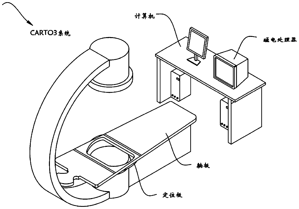
[0059] A perimeter mapping method for atrial fibrillation, such as Figure 1 to Figure 3 As shown, including a three-dimensional electroanatomical mapping system of the heart cavity and a catheter with a magnet, the method steps are as follows:

[0060] S1. Catheter positioning: After the catheter with magnet enters the magnetic field, the magnetic field processor locates the catheter by sensing the position of the magnet; (not completely correct, you can check the positioning principle of the Carto and EnSite systems)

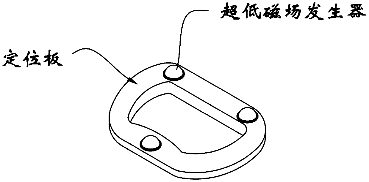
[0061] S2. Imaging modeling: The patient lies flat on the operating table, and the chest cavity is aligned with the positioning plate. When the catheter with magnets moves in the heart cavity, the computer calculates the position and potential information collected by the catheter, and displays the heart on the computer. Cavity model, and quickly build electroanatomical maps, activation conduction maps, voltage maps, impedance maps, and fragmentation point maps, et...

Embodiment 2

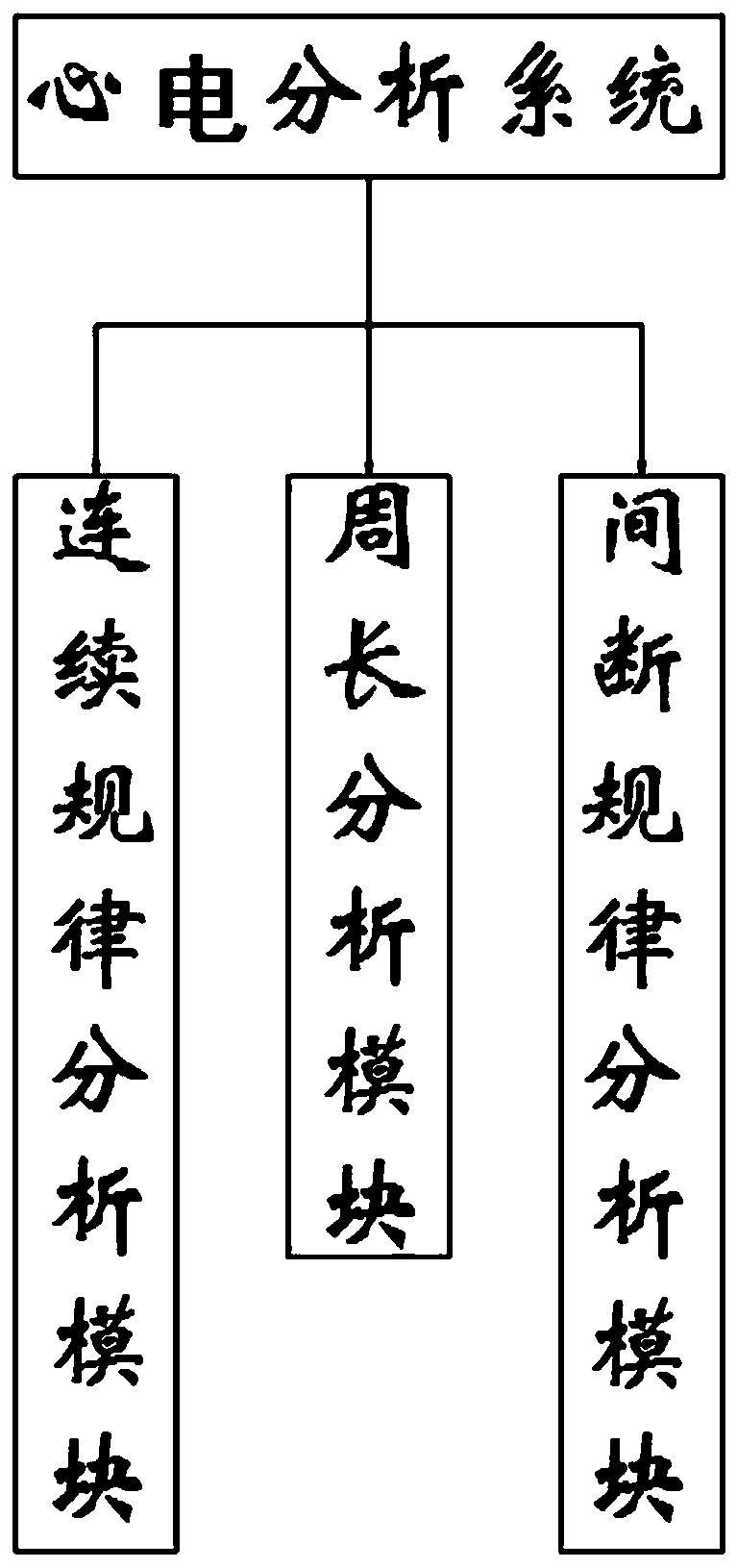
[0074] As the second embodiment of the present invention, in order to facilitate the analysis of the continuous regular heart rate, the present invention is provided with a continuous regularity analysis module, as a preferred embodiment, such as Figure 4 As shown, the ECG analysis system includes a continuous law analysis module, a circumference analysis module, and a discontinuous law analysis module. The continuous law analysis module includes a period setting module, a circumference extraction module, a circumference threshold setting module, a circumference comparison module, and The area marking module, the set period module is used to set the range of the sampling period, the circumference extraction module is used to extract the ECG perimeter within the set period, the perimeter threshold module is used to set the standard ECG perimeter , The perimeter comparison module is used to compare the ECG perimeter within the set period range with the set standard ECG perimeter, ...

Embodiment 3

[0098] As the third embodiment of the present invention, in order to facilitate the analysis of the continuous and regular heart rate circumference, the present invention is provided with a circumference analysis module, as a preferred embodiment, such as Figure 7 As shown, the perimeter analysis module includes the rule import module, the peak value extraction module, the valley value extraction module, the perimeter calculation module, the perimeter threshold setting module, the perimeter comparison module, the color distinguishing module, and the drawing and imaging module. The module is used to extract the regular excitation data obtained by the continuous law analysis module, the peak extraction module is used to determine the peak distance of the regular excitation data, the valley extraction module is used to determine the valley distance of the regular excitation data, and the circumference calculation module is used To calculate the perimeter of an excitation cycle, the...


PUM


Abstract

The invention relates to the technical field of electro cardio detection, in particular to a perimeter mapping method for auricular fibrillation. A cardiac chamber three-dimensional electro-anatomicalmapping system and a catheter with a magnet are included, and an electro cardio analysis system comprises a continuous rule analyzing module, a perimeter analyzing module and an interruption rule analyzing module. The perimeter mapping method for auricular fibrillation comprises the steps that during auricular fibrillation, atrial ordered excitation and disordered excitation portions are distinguished, during auricular fibrillation, some parts continue to be excited regularly, some parts are excited regularly or irregularly, some parts are irregular all the time, the probability that the partwhich is excited regularly at high frequency is a trigger focus is large, the parts excited regularly with the large perimeter are excited passively and are not foci, and the irregular parts with various perimeters are passively excited or are independent channels. Cardiac chamber ordered excitation and disorder excitation during auricular fibrillation are displayed on an atrial three-dimensionalmodel so as to position the source of the trigger focus, the atrial excitation conduction rules during auricular fibrillation are explained, and therefore the novel thought is supplied to radio frequency ablation during auricular fibrillation.

Description

Technical field [0001] The invention relates to the technical field of electrocardiogram detection, in particular to a perimeter mapping method for atrial fibrillation. Background technique [0002] At present, three-dimensional mapping methods for tachyarrhythmia include electroanatomical mapping, activation mapping, voltage mapping, impedance mapping, and so on. Through mapping, the activation sequence, voltage, impedance, and potential of each part of the heart cavity can be displayed on the three-dimensional anatomical model of the heart cavity, and electroanatomical maps, electrocardiograms, voltage maps, isochronous maps, pressure maps, impedance maps, etc. can be made. This helps to discover the mechanism of arrhythmia and provides guidance for catheter ablation (two mainstream mapping systems, Carto and EnSite, have these functions). These methods can meet almost all tachyarrhythmias. However, because the mechanism of atrial fibrillation is not well understood at presen...

Claims


Application Information

Patent Timeline
Patent Type & Authority Applications(China)
IPC IPC(8): A61B5/0402A61B5/0472A61B5/366
CPCA61B5/366A61B5/318
Inventor 张宏蒋建勋蒋柏涵
Owner WUXI PEOPLES HOSPITAL
Why Eureka
Try Eureka