Angle-adjustable visual uterine cavity tissue examination tube

A technology for visualizing the uterine cavity and adjusting the angle, which is applied in medical science, endoscopy, diagnosis, etc., can solve the problems of high technical requirements, anesthesia risk, and restrictions on the wide application of hysteroscopy in clinical practice, and overcome unclear vision , the effect of avoiding complications

Pending Publication Date: 2019-08-30
CHONGQING JINSHAN SCI & TECH GRP
View PDF0 Cites 0 Cited by
  • Summary
  • Abstract
  • Description
  • Claims
  • Application Information

AI Technical Summary

Problems solved by technology

[0002] At present, the clinical examination methods for gynecological intrauterine lesions and abnormalities mainly include: hysterosalpingography, X-ray hysterosalpingography, vaginal ultrasound (ultrasound), MIR and hysteroscopy. There are advantages and disadvantages in various techniques. Emphasis is also different. Hysteroscopy is comprehensive and thorough, known as the gold standard, but there are also deficiencies such as expensive equip

Method used


Image

  • Angle-adjustable visual uterine cavity tissue examination tube
  • Angle-adjustable visual uterine cavity tissue examination tube
  • Angle-adjustable visual uterine cavity tissue examination tube

Examples


Embodiment Construction

[0033] Below in conjunction with accompanying drawing, the present invention is described in detail.

[0034] In order to make the object, technical solution and advantages of the present invention clearer, the present invention will be further described in detail below in conjunction with the accompanying drawings and embodiments. It should be understood that the specific embodiments described here are only used to explain the present invention, not to limit the present invention.

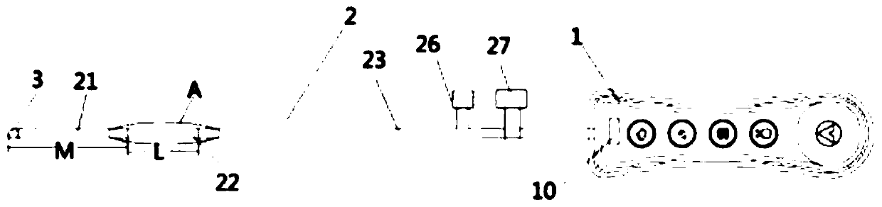
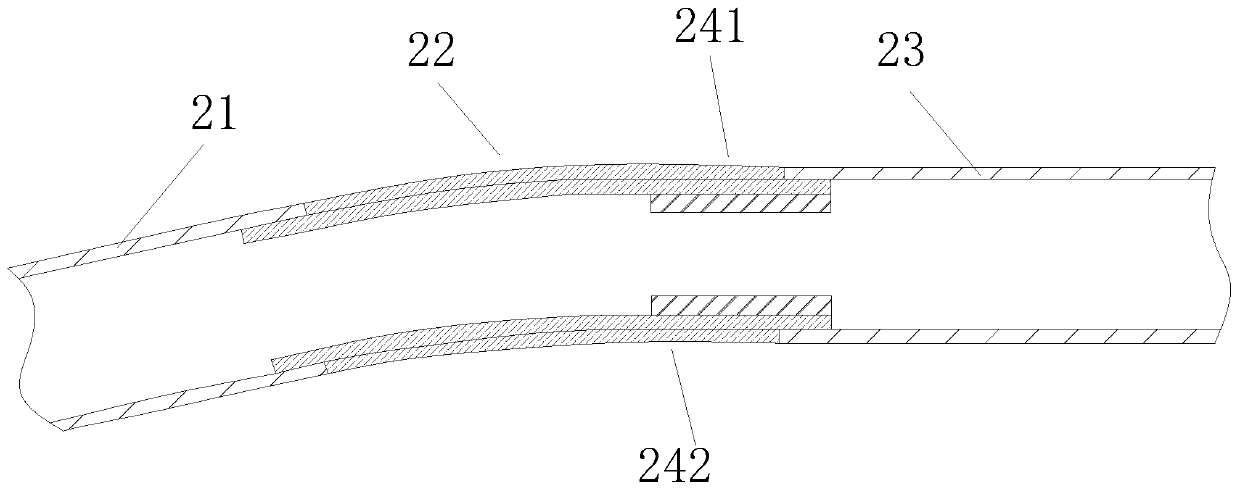
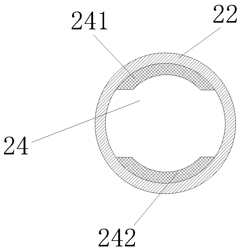
[0035] Such as Figure 1 to Figure 3 As shown, the angle-adjustable visible uterine cavity tissue examination tube provided by the present application includes a tube body 2 and a handle 1, and a circuit board 8 is arranged inside the handle 1, and one end of the tube body 2 is connected to a transparent hood 3 , the other end of the tube body 2 is connected to the handle 1; the transparent hood 3 is provided with a camera and an LED light board; the electrical connection wires of the camera and ...


PUM

PropertyMeasurementUnit
Outer diameteraaaaaaaaaa

Abstract

The invention provides an angle-adjustable visual uterine cavity tissue examination tube. The angle-adjustable visual uterine cavity tissue examination tube includes a tube body and a handle, and oneend of the tube body is connected with a transparent head cover; the tube body includes a first tube body, a second tube body and a third tube body connected in turn; and the second tube body is flexible and angle-adjustable. The tube body of the examination tube is divided into the first tube body, the second tube body and the third tube body, and the second tube body is flexible and angle-adjustable, so that the angle of the tube body can be adjusted by adjusting the bending degree of the second tube body, and the situation in the uterus can be clearly observed without distending the uterus.The invention avoids the complications caused by distending the uterus required by hysteroscopy, and overcomes the problem of unclear visual field under B-mode ultrasound.

Description

technical field [0001] The invention relates to the field of medical instruments, in particular to an angle-adjustable visible uterine cavity tissue inspection tube. Background technique [0002] At present, the clinical examination methods for gynecological intrauterine lesions and abnormalities mainly include: hysterosalpingography, X-ray hysterosalpingography, vaginal ultrasound (ultrasound), MIR and hysteroscopy. There are advantages and disadvantages in various techniques. Emphasis is also different. Hysteroscopy is comprehensive and thorough, known as the gold standard, but there are also deficiencies such as expensive equipment, high fees, anesthesia risks, and high operational technical requirements. Risks such as edema, respiratory failure and even death, and the prevention of these complications is mainly through changing the medium solution clinically, but the effect is not great, so these complications also restrict the wide clinical application of hysteroscopy. ...

Claims


Application Information

Patent Timeline
IPC IPC(8): A61B1/005A61B1/04A61B1/06
CPCA61B1/0052A61B1/0055A61B1/0058A61B1/04A61B1/0684
Inventor 朱思辉田维权冯地艮陈光柳肖建明张兴洪袁建
Owner CHONGQING JINSHAN SCI & TECH GRP
Why Eureka
Try Eureka