Bone fault image reconstruction method and system

A technology of tomographic images and images, applied in the field of nuclear medicine imaging, to facilitate analysis and diagnosis, avoid tomographic acquisition, and improve efficiency

Active Publication Date: 2019-09-20
ATOMICAL MEDICAL EQUIP FO SHAN LTD +1
View PDF2 Cites 8 Cited by
  • Summary
  • Abstract
  • Description
  • Claims
  • Application Information

AI Technical Summary

Problems solved by technology

In clinical bone gamma film images, usually only a few organs and tissues such as bones and kidneys have high radioactive uptake and image contrast, so the image data is relatively sparse, and experienced physicians are able to analyze bone anatomy. This process is completed in the physician's thinking, and there is no way to realize the intuitive registration and fusion display of the bone gamma plain film image and the CT tomographic image of the corresponding part

Method used


Image

  • Bone fault image reconstruction method and system
  • Bone fault image reconstruction method and system
  • Bone fault image reconstruction method and system

Examples


Embodiment 1

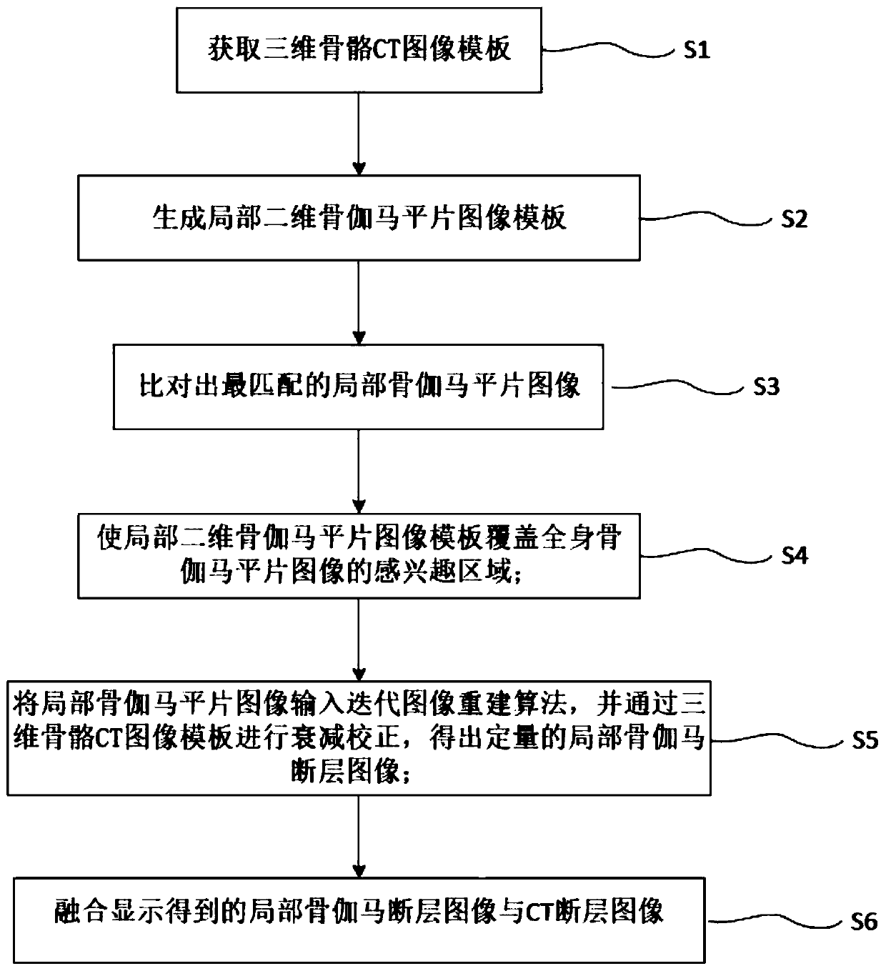
[0043] A method for reconstructing a bone tomographic image, comprising the following steps:

[0044] S1: Obtain a three-dimensional skeleton CT image template in the CT local tomographic image;

[0045] S2: Generate a local two-dimensional bone gamma plain film image template according to the three-dimensional bone CT image template;

[0046] S3: searching for the local bone gamma plain film image that best matches the local two-dimensional bone gamma plain film image template in the whole body bone gamma plain film image;

[0047]S4: According to the degree of matching between the whole-body bone gamma plain film acquisition image and the local two-dimensional bone gamma plain film image template, the size and range of the three-dimensional bone CT image template are fine-tuned using image morphology algorithms to make the local two-dimensional bone gamma The plain film image template covers the region of interest of the whole body bone gamma plain film image;

[0048] S5:...

Embodiment 2

[0061] A bone tomographic image reconstruction system, including a three-dimensional bone CT image template acquisition module, a two-dimensional bone gamma plain film image template generation module, a local bone gamma plain film image matching module, a region of interest coverage module, a reconstruction attenuation module and a fusion module;

[0062] Three-dimensional bone CT image template acquisition module: used to acquire the three-dimensional bone CT image template in the CT local tomographic image;

[0063] Two-dimensional bone gamma plain film image template generation module: used to generate a local two-dimensional bone gamma plain film image template according to the three-dimensional bone CT image template;

[0064] Local bone gamma plain film image matching module: used to search for the local bone gamma plain film image that best matches the local two-dimensional bone gamma plain film image template in the whole body bone gamma plain film image;

[0065] Re...


PUM


Abstract

The invention discloses a bone fault image reconstruction method and system. The method comprises the following steps of S1, obtaining a three-dimensional bone CT image template; S2, generating a local two-dimensional bone gamma plain film image template; S3, matching the whole body bone gamma plain film image to obtain the most matched local two-dimensional bone gamma plain film image; S4, further optimizing the three-dimensional bone CT image template, and enabling the two-dimensional bone gamma image template corresponding to the three-dimensional bone CT image template to completely cover the region of interest of the local bone gamma image; s5, reconstructing a quantitative local bone gamma tomography image based on the template; and S6, carrying out fusion display on the local bone gamma tomography image and the CT tomography image. By means of the method and system, the level close to the quality of a tomographic image obtained through conventional SPECT tomographic acquisition and reconstruction can be achieved after fusion, a doctor can analyze and diagnose the tomographic image more visually, clinical examination is conducted on part of patients, extra tomographic acquisition is avoided, and efficiency is improved.

Description

technical field [0001] The invention relates to the technical field of nuclear medicine imaging, in particular to a method and system for reconstructing a bone tomographic image. Background technique [0002] Clinically, when using SPECT (Single-Photon Emission Computed Tomography, Single-Photon Emission Computed Tomography) to examine related diseases such as malignant tumor bone metastasis, bone injury, and bone inflammation, a whole-body bone scan procedure is usually performed to obtain whole-body bone gamma Anterior and posterior plain film images are read and analyzed by physicians. For some patients with complicated conditions, it may be necessary for the physician to further decide to perform a local tomographic X-ray CT scan based on the whole-body bone gamma plain film image, and obtain a CT tomographic image of a certain part for auxiliary diagnosis. If there is still doubt about the combination of bone plain film images and CT local tomographic images, SPECT tom...

Claims


Application Information

Patent Timeline
Patent Type & Authority Applications(China)
IPC IPC(8): G06T17/00G06T7/11G06T11/00
CPCG06T11/003G06T17/00G06T2207/10081G06T2207/30008G06T7/11
Inventor 陈思姜苏青蔡奉岐
Owner ATOMICAL MEDICAL EQUIP FO SHAN LTD
Why Eureka
Try Eureka