
Automatic organ-at-risk sketching method and device based on neural network and storage medium

A neural network, organ technology

Active Publication Date: 2019-10-08
BEIJING LINKING MEDICAL TECH CO LTD
View PDF9 Cites 20 Cited by
  • Summary
  • Abstract
  • Description
  • Claims
  • Application Information

AI Technical Summary

Problems solved by technology

Due to the characteristics of fast imaging speed, high spatial accuracy and high resolution, CT images are usually used to formulate radiotherapy plans. Doctors need to accurately delineate each organ at risk in each CT slice, which is a time-consuming and laborious process. In addition, , due to the low contrast of soft tissues in CT images, for example, the parotid gland has no clear boundary and complex shape, which makes it error-prone and time-consuming for doctors to draw manually, so an accurate and fast automatic organ-at-risk segmentation algorithm is needed to assist Physicians delineate organs at risk, reducing physical labor and time in the planning process
[0003] The current products on the market are all based on the Multi-Atlas (multi-atlas) registration method. The segmentation accuracy of this method depends on the selection of the template, which is less robust and cannot adapt to CT with different resolutions in different hospitals. Image data, and, due to the use of deformation registration, the segmentation time is longer
On the other hand, in order to improve the accuracy of segmentation, as many templates as possible will be selected, but the segmentation time will also increase with the increase of templates, and many current methods use a lot of prior knowledge, and the generalization ability is poor.

Method used


Image

  • Automatic organ-at-risk sketching method and device based on neural network and storage medium
  • Automatic organ-at-risk sketching method and device based on neural network and storage medium
  • Automatic organ-at-risk sketching method and device based on neural network and storage medium

Examples


Embodiment 2

[0122] The invention also provides a computing device, comprising:

[0123] one or more processors;

[0124] storage; and

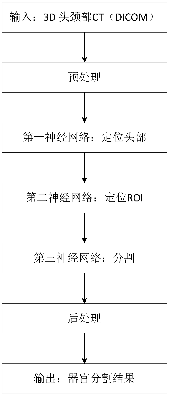
[0125] One or more programs, wherein the one or more programs are stored in the memory and configured to be executed by the one or more processors, the one or more programs include the above-mentioned three-cascade volume-based Instructions for the method for automatically delineating organs at risk by the product neural network, the method includes the following steps:

[0126] (1) Input 3D medical images;

[0127] (2) Preprocessing the 3D medical image;

[0128] (3) Input the preprocessed three-dimensional medical image into the first-level network of the trained three-cascade convolutional neural network to identify the cross-section of the organ to be segmented;

[0129] (4) Input the cross-section screened by the first-level network into the second-level network of the trained three-cascade convolutional neural network, and roughly locate the reg...

Embodiment 3

[0135] The present invention also provides a computer-readable storage medium that stores one or more programs, and the one or more programs include instructions, and the instructions are suitable for being loaded by the memory and executing the above-mentioned three-cascade convolutional neural network-based A method for automatically delineating organs at risk, the method comprising the following steps:

[0136] (1) Input 3D medical images;

[0137] (2) Preprocessing the 3D medical image;

[0138] (3) Input the preprocessed three-dimensional medical image into the first-level network of the trained three-cascade convolutional neural network to identify the cross-section of the organ to be segmented;

[0139] (4) Input the cross-section screened by the first-level network into the second-level network of the trained three-cascade convolutional neural network, and roughly locate the region of interest of the organ to be segmented;

[0140] (5) Standardize the region of inter...


PUM


Abstract

The invention belongs to the technical field of medical images, and relates to an automatic organ-at-risk sketching method and device based on a three-level convolutional neural network, and a storagemedium. The method comprises the following steps of: preprocessing the three-dimensional medical image, inputting the preprocessed three-dimensional medical image into the first-stage network, the second-stage network and the third-stage network of the trained three-stage convolutional neural network, sequentially identifying the cross section of the organ to be segmented, coarsely positioning the region of interest of the organ to be segmented, and classifying all pixel points in the region of interest; and then outputting a three-dimensional binary segmentation result; carrying out post-processing, edge extraction and edge smoothing on the binary segmentation result to obtain an automatically sketched organ. The three-level cascade convolutional neural network model is formed by cascading three convolutional neural networks, namely a first-level network, a second-level network and a third-level network. The three-level joint neural network has the advantages that priori knowledge isnot needed, the algorithm generalization ability is good, the robustness is high, the speed is high, full automation is achieved, and the segmentation accuracy is high.

Description

technical field [0001] The invention belongs to the field of medical imaging and computer technology, and relates to a method, device and storage medium for automatically delineating organs at risk based on a three-cascade convolutional neural network. Background technique [0002] Radiation therapy is one of the three major methods of cancer treatment. It can destroy the DNA chain of cancer cells through ionizing radiation, and then lead to the death of cancer cells. In order to reduce the impact of radiation on normal tissues during treatment, doctors need to make a careful radiotherapy plan before radiotherapy. Due to the characteristics of fast imaging speed, high spatial accuracy and high resolution, CT images are usually used to formulate radiotherapy plans. Doctors need to accurately delineate each organ at risk in each CT slice, which is a time-consuming and laborious process. In addition, , due to the low contrast of soft tissues in CT images, for example, the paro...

Claims


Application Information

Patent Timeline
IPC IPC(8): G06T7/11G06T7/13G06T15/00G06N3/04G06N3/08G06K9/32
CPCG06T7/11G06T7/13G06T15/005G06N3/084G06T2207/10081G06T2207/30016G06V10/25G06N3/048G06N3/045
Inventor 孙鑫龙崔德琪章桦
Owner BEIJING LINKING MEDICAL TECH CO LTD
Learn More