
Medical image quality evaluation method, device, equipment and storage medium

An image quality assessment and image quality technology, applied in the field of medical image processing, can solve the problems of increasing the workload of doctors, time-consuming and labor-intensive problems, and achieve the effect of avoiding inaccurate follow-up diagnosis and reducing workload

Active Publication Date: 2019-11-08
SHANGHAI UNITED IMAGING HEALTHCARE
View PDF17 Cites 11 Cited by
  • Summary
  • Abstract
  • Description
  • Claims
  • Application Information

AI Technical Summary

Problems solved by technology

However, each subject may correspond to multiple reconstructed images. Take the whole body MRI scan as an example. On the one hand, due to the limitation of scanning hardware, the whole body scan can only be completed by scanning several beds separately; on the other hand, each A routine MRI scan on a bed should include images with different weights such as T1, T2, and DWI, and images with the same weight should include different acquisition orientations such as T2 transverse and T2 coronal, acquisitions with different parameters, and / or, some diseases will More targeted MRI scan sequences are added in some parts, therefore, there may be at least twenty MRI scan sequences, that is, there may be at least twenty reconstructed images for each subject, which allows physicians to observe the images The quality process is quite time-consuming and laborious, increasing the workload of physicians

Method used


Image

  • Medical image quality evaluation method, device, equipment and storage medium
  • Medical image quality evaluation method, device, equipment and storage medium
  • Medical image quality evaluation method, device, equipment and storage medium

Examples


Embodiment 1

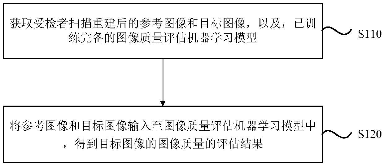
[0046] figure 1 It is a flowchart of a medical image quality assessment method provided in Embodiment 1 of the present invention. This embodiment is applicable to the situation of evaluating the quality of medical images after scanning and reconstruction, and is especially suitable for evaluating whether there is a corresponding image in the target image after scanning and reconstruction by using a reference image without image quality defects to be evaluated as the gold standard In case of quality defects. The method can be executed by the medical image quality assessment device provided by the embodiment of the present invention, the device can be realized by software and / or hardware, and the device can be integrated on various devices.

[0047] see figure 1 , the method of the embodiment of the present invention specifically includes the following steps:

[0048] S110. Acquire the scanned and reconstructed reference image and the target image of the subject, and a fully ...

Embodiment 2

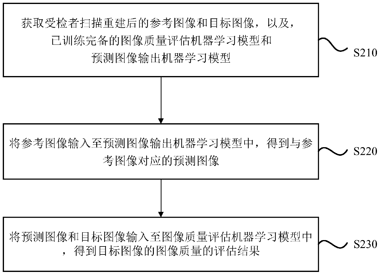
[0062] figure 2 It is a flowchart of a medical image quality assessment method provided in Embodiment 2 of the present invention. This embodiment is optimized on the basis of the above-mentioned technical solutions. In this embodiment, optionally, the medical image quality assessment method may specifically include: obtaining a fully trained predictive image output machine learning model; correspondingly, inputting the reference image and the target image into the image quality assessment machine In the learning model, obtaining the evaluation result of the image quality of the target image may include: inputting the reference image into the predicted image output machine learning model to obtain a predicted image corresponding to the reference image; inputting the predicted image and the target image into the image quality assessment In the machine learning model, the evaluation result of the image quality of the target image is obtained. Wherein, explanations of terms tha...

Embodiment 3

[0078] Figure 5 It is a structural block diagram of a medical image quality assessment device provided in Embodiment 3 of the present invention, and the device is used to implement the medical image quality assessment method provided in any of the above-mentioned embodiments. The device and the medical image quality assessment method of the above-mentioned embodiments belong to the same inventive concept. For details not described in detail in the embodiments of the medical image quality assessment device, you can refer to the above-mentioned embodiment of the medical image quality assessment method . see Figure 5 , the device may specifically include: an acquisition module 310 and an image quality evaluation module 320 .

[0079] Wherein, the obtaining module 310 is used to obtain the reference image and the target image after scanning and reconstruction of the subject, and a fully trained image quality assessment machine learning model, wherein the reference image and the ...


PUM


Abstract

The embodiment of the invention discloses a medical image quality evaluation method, a device, equipment and a storage medium. The method comprises the following steps: acquiring a reference image anda target image which are scanned and reconstructed by a subject and a trained complete image quality evaluation machine learning model, the reference image and the target image have the same fault information and position information, and the reference image does not have image quality defects to be evaluated corresponding to the image quality evaluation machine learning model; and inputting thereference image and the target image into an image quality evaluation machine learning model to obtain an evaluation result of the image quality of the target image. According to the medical image quality evaluation method, by taking the reference image without the to-be-evaluated image quality defect as a gold standard, whether the target image has the image quality defect is evaluated, so that adoctor can make a scanning decision according to a quality evaluation result, the workload of the doctor can be effectively reduced, and subsequent diagnosis is prevented from being influenced by a medical image with poor quality.

Description

technical field [0001] Embodiments of the present invention relate to the technical field of medical image processing, and in particular, to a method, device, device, and storage medium for evaluating the quality of medical images. Background technique [0002] In medical imaging systems, the image quality depends on many factors, such as spatial resolution, tissue contrast, signal-to-noise ratio, etc. The defects in image quality caused by these factors can be improved by optimizing hardware and scanning parameters. This results in better image quality; however, in some cases, for example, the defect of metal artifacts caused by metal devices such as surgical instruments, motion artifacts caused by the subject's movement during the scanning process such as breathing, heartbeat, etc. The defect of imaging is that better image quality cannot be obtained through optimization of hardware and scanning parameters, and poor image quality cannot meet the requirements of clinical di...

Claims


Application Information

Patent Timeline
IPC IPC(8): G06T7/00G06N20/00G06N3/08G06N3/04
CPCG06T7/0012G06N3/08G06N20/00G06T2207/30168G06N3/044
Inventor 史宇航
Owner SHANGHAI UNITED IMAGING HEALTHCARE
Learn More