Automatic measuring method and device of intrapartum cephalopelvic relationship based on ultrasonoscopy

An ultrasonic image, automatic measurement technology, applied in ultrasonic/sonic/infrasound image/data processing, ultrasonic/sonic/infrasonic Permian technology, organ motion/change detection, etc., can solve the problem of missing edges and poor real-time performance , difficult to apply and other problems, to achieve the effect of reducing computational overhead, improving sensitivity and prediction accuracy, and reducing the rate of cesarean section

Inactive Publication Date: 2019-11-12
JINAN UNIVERSITY
View PDF8 Cites 6 Cited by
  • Summary
  • Abstract
  • Description
  • Claims
  • Application Information

AI Technical Summary

Problems solved by technology

However, due to the inconspicuous features of the pubic symphysis in ultrasound images, large morphological differences, severe edge loss, and serious edge loss of the fetal head contour, it is very difficult to realize the automatic measurement of intrapartum fetal ultrasound parameters. Therefore, the current intrapartum fetal ultrasound parameters There are few related studies on automatic measurement, and most of them stay at the theoretical stage, with poor real-time performance and difficult to apply clinically
Moreover, the existing measurement methods all need manual marking for assistance or use prior knowledge for measurement, which has limitations, and there is no automatic measurement method without manual intervention

Method used


Image

  • Automatic measuring method and device of intrapartum cephalopelvic relationship based on ultrasonoscopy
  • Automatic measuring method and device of intrapartum cephalopelvic relationship based on ultrasonoscopy
  • Automatic measuring method and device of intrapartum cephalopelvic relationship based on ultrasonoscopy

Examples


Embodiment 1

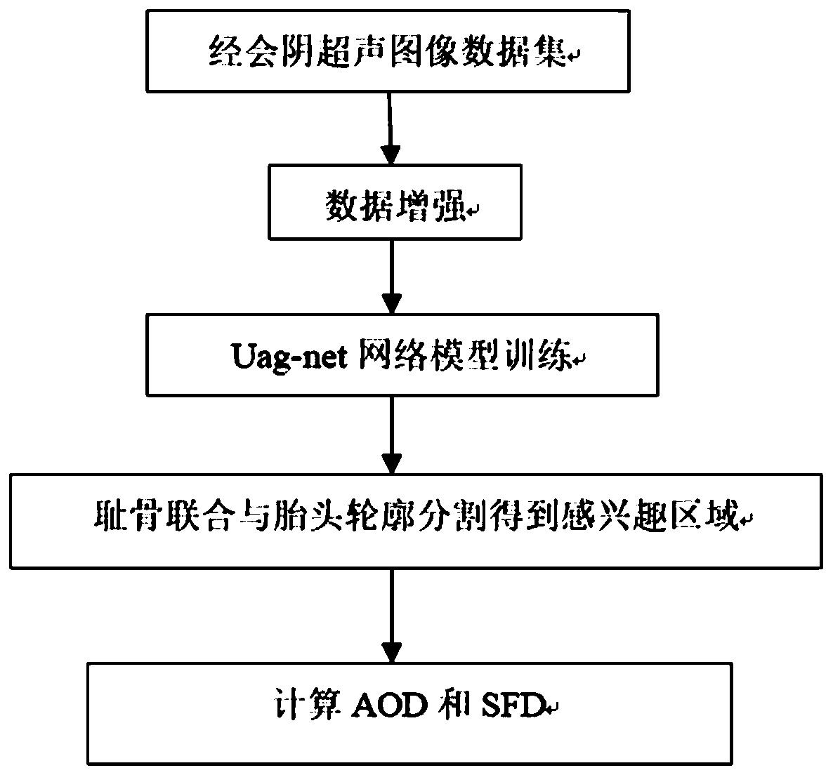
[0042] Such as figure 1 As shown, a method for automatic measurement of head-pelvic relationship based on ultrasound images mainly includes the following steps:

[0043] S1. Obtain a maternal transperineal ultrasound image data set for training, perform data enhancement on the intrapartum transperineal ultrasound image data set, and obtain the enhanced data set as a training set;

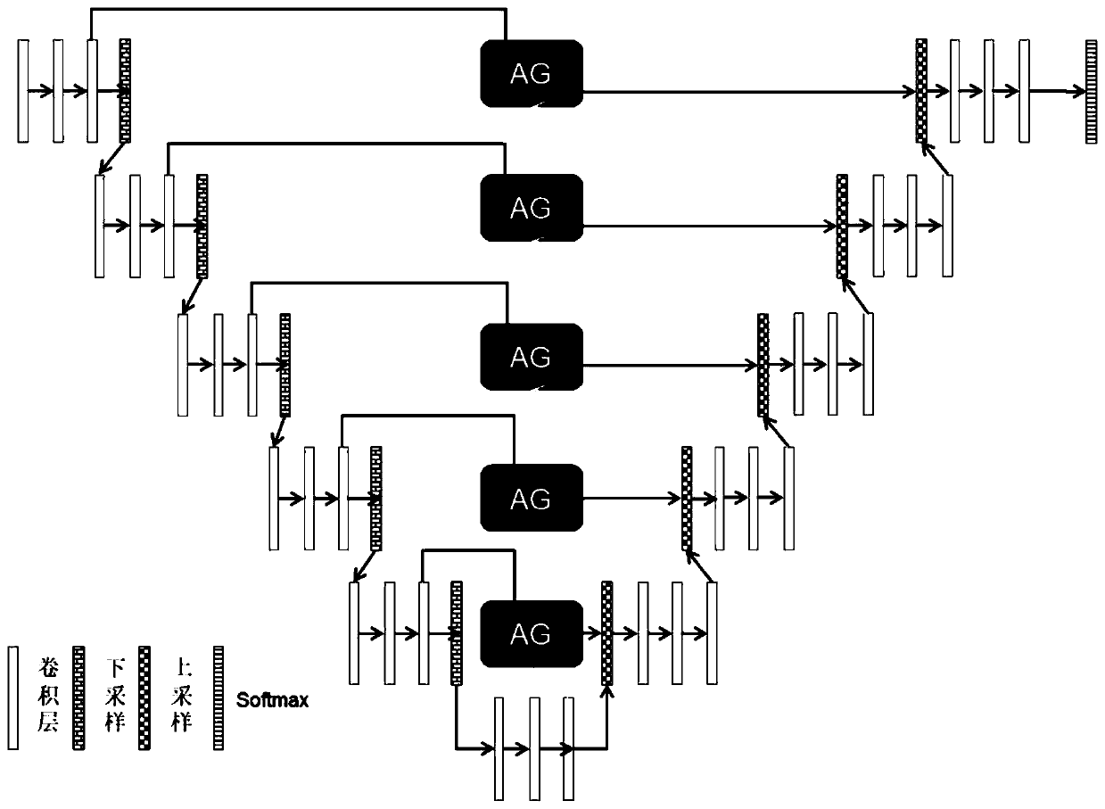
[0044] S2. Input the data in the training set into the constructed neural network model Uag-net, train the end-to-end segmentation model, and segment the region of interest of the pubic symphysis and the region of interest of the fetal head; train repeatedly to obtain a well-trained neural network model;

[0045] S3. In actual application, input the real-time acquired ultrasound image into the trained neural network model to obtain the region of interest of the pubic symphysis and the region of interest of the fetal head, and perform enhancement and boundary fitting on the region of interest, Cal...

Embodiment 2

[0078] Present embodiment except following feature other structures are with embodiment 1:

[0079] An automatic measurement device for head-pelvic relationship based on ultrasound images, comprising:

[0080] The training set building block is used to obtain the maternal transperineal ultrasound image data set used for training as a training set;

[0081] The neural network model training module is used to train an end-to-end segmentation model according to the data in the training set, and segment the region of interest of the pubic symphysis and the region of interest of the fetal head; repeated training to obtain a trained neural network model;

[0082] The parameter calculation module is used to input the real-time collected ultrasound images into the trained neural network model to obtain the region of interest of the pubic symphysis and the region of interest of the fetal head, enhance and fit the region of interest, and calculate Head-pelvis relationship parameters, the...


PUM


Abstract

The invention discloses an automatic measuring method and device of intrapartum cephalopelvic relationship based on ultrasonoscopy. The automatic measuring method comprises the steps that a puerpera transperineal ultrasonoscopy data set for training is obtained, and taken as a training set; data in the training set are input into a constructed neural network model, an end-to-end split model is trained, and interested areas of the symphysis pubis and the fetus head are split; training is conducted repeatedly, a trained neural network model is obtained; and in practice application, the ultrasonoscopy collected in real time is input into the trained neural network model, the interested areas of the symphysis pubis and the fetus head are obtained, enhancing and edging fitting are conducted onthe interested areas, and the fetus head angle of descending AOD of and the symphysis pubis-fetus head distance SFD are calculated. Compared with an existing intrapartum fetus parameter measuring method, according to the automatic measuring method and device, intrapartum fetus parameters can be accurately and objectively measured in real time, any priori knowledge and human intervention are not needed, the requirement of real-time application can be met, and clinical application prospects are achieved.

Description

technical field [0001] The invention belongs to the field of computer vision aided diagnosis, and in particular relates to the research on the segmentation method of transperineal ultrasonic images during labor, in particular to a method and device for automatically measuring the head-pelvic relationship during labor based on ultrasonic images. Background technique [0002] Factors affecting childbirth include four factors: labor force, birth canal, fetus, and mental psychology. The normal head delivery mechanism can be described as: when the fetal part passes through the birth canal, in order to adapt to the different shapes of the different planes of the pelvis, it performs a passive movement. A series of adaptive rotations, the process of passing through the birth canal with its smallest diameter. It can be seen that the core of the delivery mechanism is the lowering of the fetal head, and the intuitive description of head-position delivery is the movement trajectory of t...

Claims


Application Information

Patent Timeline
Patent Type & Authority Applications(China)
IPC IPC(8): A61B8/08
CPCA61B8/0866A61B8/5215
Inventor 陆尧胜周铭鸿袁超齐建国
Owner JINAN UNIVERSITY
Why Eureka
Try Eureka