Endoscope image imaging method, device, apparatus and medium

An endoscope and image technology, applied in the field of medical detection, can solve the problems of uneven image quality, influence, and the inability of traditional algorithms to complete image enhancement, etc., to achieve the effect of enhancing contrast

Inactive Publication Date: 2019-12-03
SUN YAT SEN UNIV
View PDF3 Cites 5 Cited by
  • Summary
  • Abstract
  • Description
  • Claims
  • Application Information

AI Technical Summary

Problems solved by technology

[0003] However, the shooting of endoscopic images is affected by gas bubbles in the digestive tract, light and shooting angles, resulting in uneven image quality. The complexity of the environment structure - the impact of gas bubbles in the digestive tract, etc., the image reflected by the endoscope is not ideal, and the large area of ​​the image is dark due to underexposure, which has a great impact on the evaluation of the patient's disease during the operation
[0004] There are many traditional image enhancement methods, such as histogram equalization enhancement, Laplace operator enhancement, gamma transformation enhancement, and multi-scale image enhancement algorithms. Due to the special scene of vascular endoscope, the illumination and angle are different. Traditional algorithms cannot complete image enhancement and are not suitable for this scene

Method used


Image

  • Endoscope image imaging method, device, apparatus and medium
  • Endoscope image imaging method, device, apparatus and medium
  • Endoscope image imaging method, device, apparatus and medium

Examples


Embodiment Construction

[0043] In order to make the purpose, features and advantages of the present application more obvious and understandable, the present application will be further described in detail below in conjunction with the accompanying drawings and specific implementation methods. Apparently, the described embodiments are some of the embodiments of the present application, but not all of them. Based on the embodiments in this application, all other embodiments obtained by persons of ordinary skill in the art without creative efforts fall within the protection scope of this application.

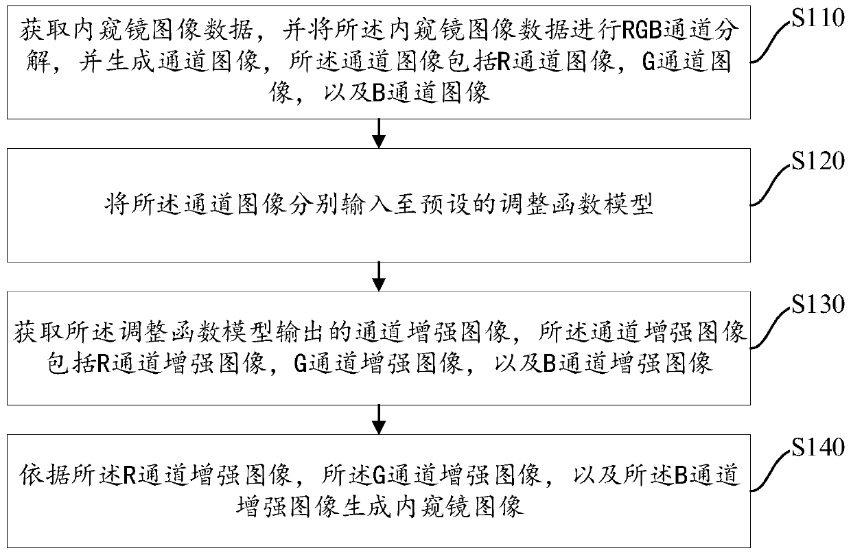
[0044] It should be noted that, in any embodiment of the present invention, the imaging method is applied to distinguish the tissue background and blood vessel features in the endoscopic image, so as to improve the accuracy of doctor's interpretation.

[0045] It should be noted that, since the surface of the oral cavity, esophagus, stomach, intestines, urethra and other organs of the human body is compos...


PUM


Abstract

The invention provides an endoscope image imaging method, device and apparatus and a medium. The method relates to adjustment of channel images of R, G and B channel components in an endoscopic image,and comprises the following steps: acquiring endoscopic image data, performing RGB channel decomposition on the endoscopic image data, and generating channel images, the channel images comprising anR channel image, a G channel image and a B channel image; respectively inputting the channel images into a preset adjustment function model; obtaining a channel enhanced image output by the adjustmentfunction model, wherein the channel enhanced image comprises an R channel enhanced image, a G channel enhanced image and a B channel enhanced image; and generating an endoscope image according to theR channel enhanced image, the G channel enhanced image and the B channel enhanced image. R, G and B channel components of an image are independently adjusted, so that an obvious contrast effect is generated between a tissue background and the hue of a blood vessel.

Description

technical field [0001] The present application relates to the field of medical detection, in particular to imaging methods, devices, equipment and media of endoscopic images. Background technique [0002] An endoscope is an optical device that can detect the internal image information of an object. Medical endoscopes can enter the human body through the natural channels of the human body or small surgical incisions, and can help doctors see the internal structure of the human body that cannot be displayed by X-rays. It has become an indispensable instrument for checking internal diseases of the stomach, esophagus, and small intestine. Obtaining clear images through the endoscope can help doctors establish patient information, which can effectively improve the accuracy of the diagnosis and treatment process. [0003] However, the shooting of endoscopic images is affected by gas bubbles in the digestive tract, light and shooting angles, resulting in uneven image quality. Due...

Claims


Application Information

Patent Timeline
Patent Type & Authority Applications(China)
IPC IPC(8): G06T5/00G16H30/40A61B1/00
CPCG06T5/008G06T5/002G16H30/40A61B1/00009G06T2207/10068G06T2207/30101
Inventor 张贺晔李吉平吴万庆
Owner SUN YAT SEN UNIV
Why Eureka
Try Eureka