An electrochemical immunosensor for detecting secreted autophagosomes and its preparation method and application
An immune sensor and autophagosome technology, which is applied in the field of biomedical detection, can solve the problems of rapid, simple and direct detection of secreted autophagosomes, and achieve excellent anti-interference performance and high selectivity.
- Summary
- Abstract
- Description
- Claims
- Application Information
AI Technical Summary
Problems solved by technology
Method used
Image
Examples
Embodiment 1
[0058] Example 1: Fe 3 o 4 - Preparation of Au Secondary Antibody Nanomagnetic Beads
[0059] (1) Fe 3 o 4 preparation of
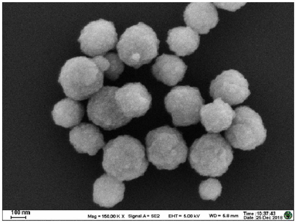
[0060] 0.65g FeCl 3 Dissolve 0.2g of sodium citrate in 20mL of ethylene glycol, then add 1.2g of sodium acetate, stir vigorously for 30 minutes, pour into a hydrothermal reactor, and react at 200°C for 10 hours. After the end of the reaction, the material is centrifuged to obtain the first precipitate. After the first precipitate is washed with water, it is centrifuged to obtain the second precipitate, and after the second precipitate is washed with ethanol, a liquid dissolved in ethanol is obtained to obtain Fe 3 o 4 (Concentration is 1mg / mL, pH value is 9.5), and its scanning electron microscope picture is as follows figure 1 shown.
[0061] (2) Preparation of AuNPs
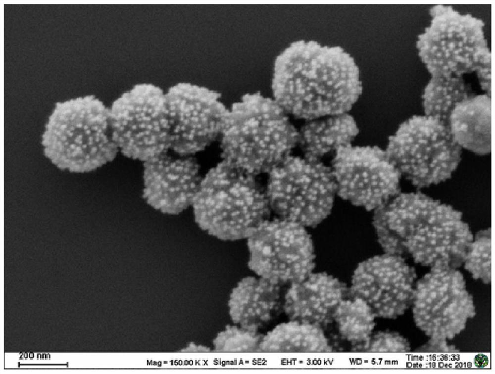
[0062] 100mL mass fraction 0.01% HAuCl 4 The aqueous solution was heated to boiling, and 2 mL of a 1% sodium citrate aqueous solution was added with stirring, and continued heati...
Embodiment 2
[0067] Example 2: Fe 3 o 4 -Specific verification of Au secondary antibody nano-magnetic beads
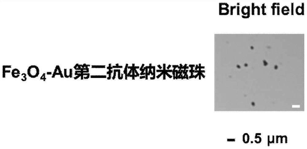
[0068] Fe 3 o 4 - Au secondary antibody nano-magnetic beads and interference red fluorescent protein co-incubated at room temperature for 2 hours, another group of Fe 3 o 4 -Au secondary antibody nano-magnetic beads were incubated with red fluorescent protein and TRAP stained with green fluorescence for 2 hours at room temperature, magnetically separated, washed 3 times in PBS, and resuspended in PBS. It was found that nano-magnetic beads can specifically capture TRAP, but not Non-specific adsorption of interfering proteins. Laser confocal images such as Figure 4 shown.
Embodiment 3
[0069] Example 3: Preparation method and detection method of electrochemical immunosensor for measuring secreted autophagosomes
[0070] The preparation method of electrochemical immunosensor is as follows: Figure 5 shown, including the following steps:
[0071] (1) Electrode pretreatment: use 0.05 and 0.03 μm Al on glassy carbon electrodes respectively 2 o 3 Powder treatment, and then ultrasonic cleaning with absolute ethanol and ultrapure water for 5 minutes;
[0072] (2) Adding GH-MB dropwise: Take 10 μL of 1 mg / mL GH-MB solution dropwise on the surface of the pretreated glassy carbon electrode with a pipette gun, place it at 37°C for 2 hours to dry, wash the unbound composite material with PBS solution, and let it dry in the air. Dry, take advantage of its larger specific surface area to enrich more antibodies on the electrode surface;
[0073] (3) Covalently link the primary antibody to the capture antibody LC3B: Add 10 μL of DC / NHS solution (20 mg / mL, 10 mg / mL) drop...
PUM
| Property | Measurement | Unit |
|---|---|---|
| concentration | aaaaa | aaaaa |
| concentration | aaaaa | aaaaa |
| concentration | aaaaa | aaaaa |
Abstract
Description
Claims
Application Information
 - R&D
- Intellectual Property
- Life Sciences
- Materials
- Tech Scout
- Unparalleled Data Quality
- Higher Quality Content
- 60% Fewer Hallucinations
Browse by: Latest US Patents, China's latest patents, Technical Efficacy Thesaurus, Application Domain, Technology Topic, Popular Technical Reports.
© 2025 PatSnap. All rights reserved.Legal|Privacy policy|Modern Slavery Act Transparency Statement|Sitemap|About US| Contact US: help@patsnap.com


