Label-free high-speed microscopic imaging method and device

A microscopic imaging, label-free technology, applied in the field of optical microscopy, can solve the problems of slow response speed, wasting time, reducing imaging speed and data throughput of pathological slices, etc. The effect of a quick check

Active Publication Date: 2021-03-02
TSINGHUA UNIV
View PDF8 Cites 0 Cited by
  • Summary
  • Abstract
  • Description
  • Claims
  • Application Information

AI Technical Summary

Problems solved by technology

The lateral movement of the sample usually has a slow response speed, which wastes a lot of time, further reducing the imaging speed and data throughput of pathology slides

Method used


Image

  • Label-free high-speed microscopic imaging method and device
  • Label-free high-speed microscopic imaging method and device
  • Label-free high-speed microscopic imaging method and device

Examples


Embodiment Construction

[0030] Embodiments of the present invention are described in detail below, examples of which are shown in the drawings, wherein the same or similar reference numerals designate the same or similar elements or elements having the same or similar functions throughout. The embodiments described below by referring to the figures are exemplary and are intended to explain the present invention and should not be construed as limiting the present invention.

[0031] The label-free high-speed microscopic imaging method and device according to the embodiments of the present invention will be described below with reference to the accompanying drawings. First, the label-free high-speed microscopic imaging method according to the embodiments of the present invention will be described with reference to the accompanying drawings.

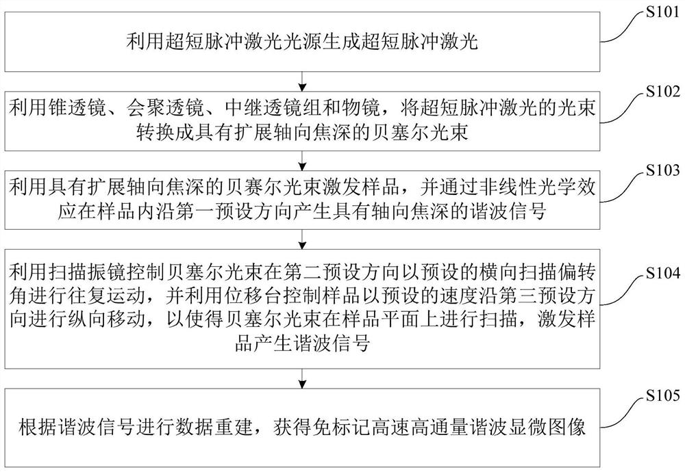
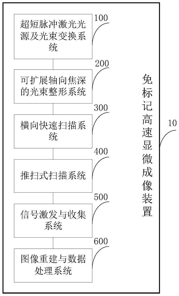
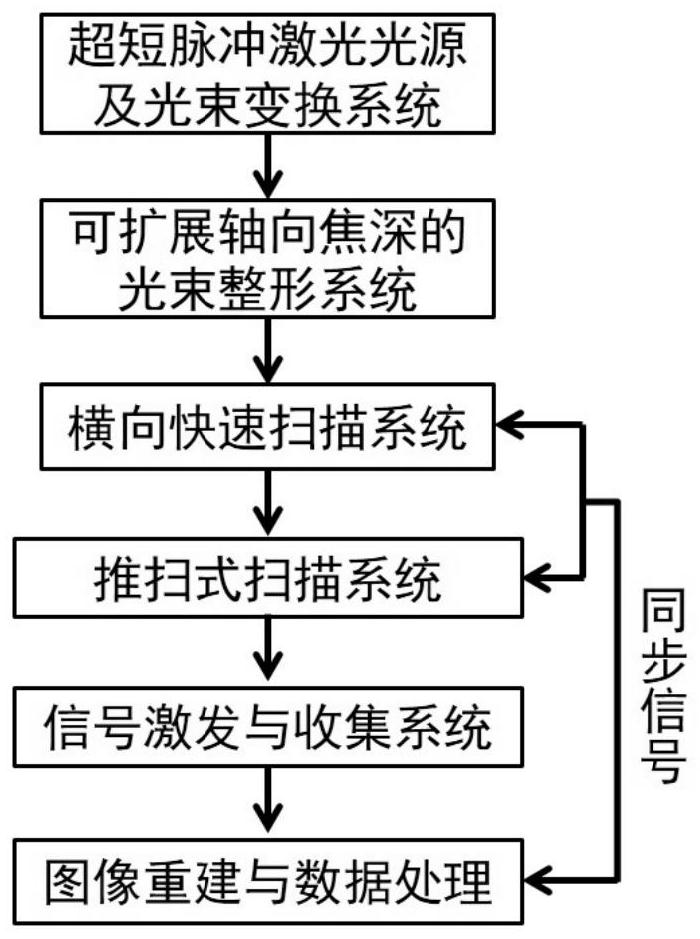
[0032] figure 1 It is a flowchart of a label-free high-speed microscopic imaging method according to an embodiment of the present invention.

[0033] Such as figu...


PUM


Abstract

The invention discloses a label-free high-speed microscopic imaging method and device. The method comprises the steps of generating ultrashort pulse laser by using an ultrashort pulse laser light source; converting a beam of the ultrashort pulse laser into a Bessel beam with an extended axial focal depth, exciting a sample by using the Bessel beam and generating a harmonic signal with an axial focal depth in the sample through a nonlinear optical effect along a first preset direction; controlling the Bessel beam to carry out reciprocating motion at a preset horizontal scanning deflection anglein a second preset direction and controlling the sample to carry out longitudinal movement at a preset speed in a third preset direction to scan the Bessel beam in a sample plane and to excite the sample to generate the harmonic signal; and carrying out data reconstruction according to the harmonic signal to obtain a label-free high-speed and high-flux harmonic microscopic image. The method is capable of significantly improving the imaging speed and the data flux, can be applied to fast imaging of biological samples, such as pathological sections, and has a broad biomedical application prospect.

Description

technical field [0001] The invention relates to the field of optical microscopy technology, in particular to a label-free high-speed microscopic imaging method and device. Background technique [0002] Three-dimensional imaging of biological samples has important applications in many biomedical researches. As a routine method of clinical examination, pathological biopsy plays an important role in clinical diagnosis. At present, traditional hematoxylin-eosin staining (H&E staining) is usually used to stain biopsy sections in pathological biopsy, which is regarded as the "gold standard" for pathological detection. However, the H&E staining method requires complex pretreatment processes such as fixation, dehydration, wax immersion, dewaxing, sectioning, and staining, and then placing the sample under a microscope for observation and imaging, which cannot achieve real-time observation and diagnosis. In order to improve the efficiency of pathological detection and avoid the abo...

Claims


Application Information

Patent Timeline
Patent Type & Authority Patents(China)
IPC IPC(8): G01N21/84G01N21/01G02B21/00
CPCG01N21/01G01N21/84G01N2021/0112G02B21/0048G02B21/0052
Inventor 孔令杰靳程戴琼海
Owner TSINGHUA UNIV
Why Eureka
Try Eureka