
Microfluidic integrated microarrays for biological detection

a microfluidic chip and integrated technology, applied in the field of microfluidic devices, can solve the problems of long and tedious processing time of rna/dna sample preparation, affecting the detection efficiency of microfluidic chips, and requiring up to four days, so as to reduce the required sample volume, reduce the preparation time of samples, and reduce the effect of required sample volum

Inactive Publication Date: 2005-05-05
SANDIA NAT LAB
View PDF32 Cites 124 Cited by
  • Summary
  • Abstract
  • Description
  • Claims
  • Application Information

AI Technical Summary

Benefits of technology

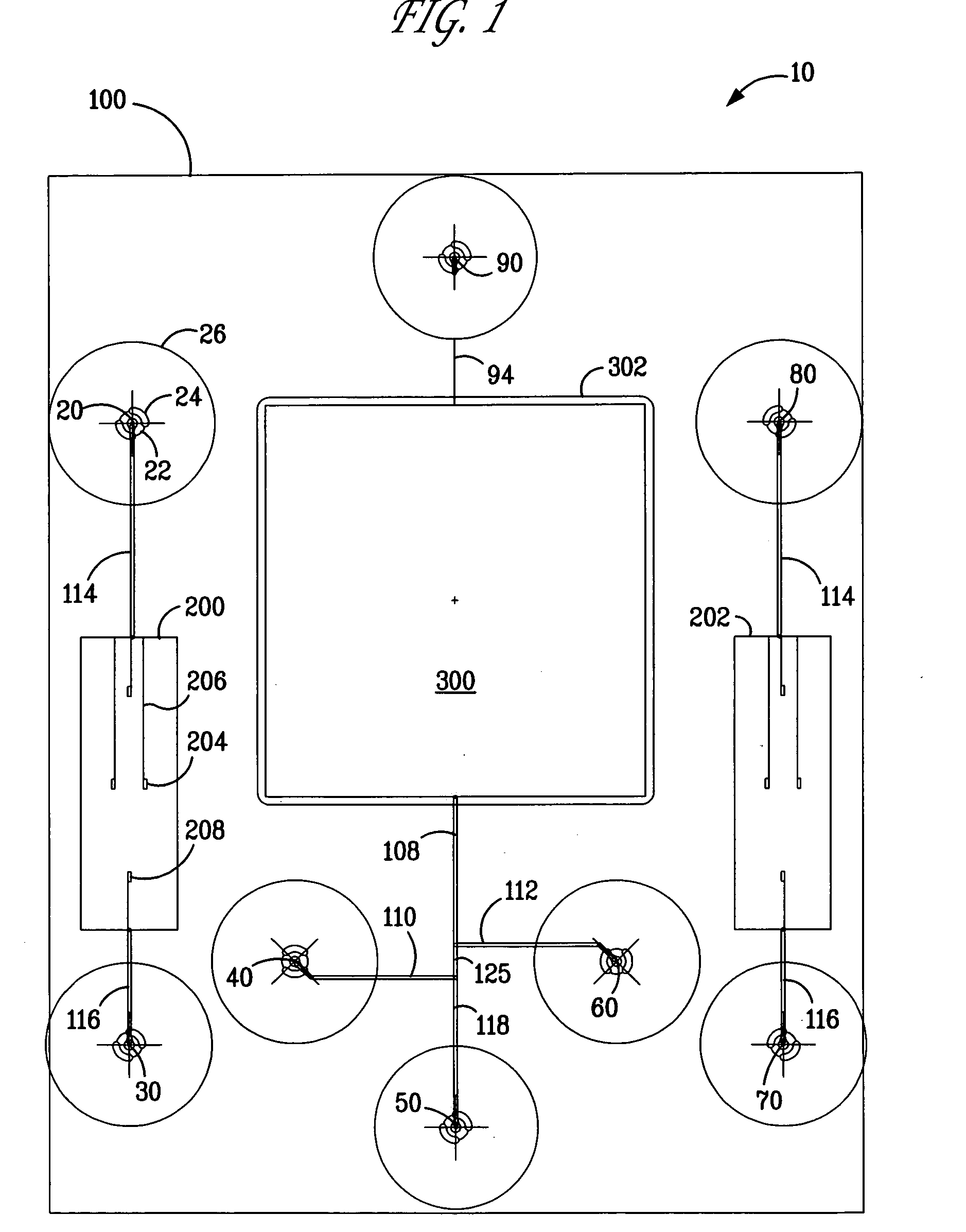
[0008] In overcoming the problems associated with providing a high throughput microfluidic chip capable of specifically capturing and concentrating nucleic acids for microarray analysis, the present invention provides, inter alia, microfluidic chips containing functionalized porous polymer monoliths for capturing and concentrating sample nucleic acids. In one aspect of the present invention, there are provided microfluidic chips that include a plurality of vias; a functionalized porous polymer monolith capable of being in fluid communication with a via; a microarray capable of being in fluid communication with the functionalized porous polymer monolith; and an observation port through which at least one target disposed within the microarray is capable of being detected. As will be disclosed in further detail below, the microfluidic chips of the present invention are capable of capturing and concentrating genetic material for the analysis and identification of biological organisms, such as the so-called “threat genes” from biological weapons. The microarrays are capable of being in fluid communication with the functionalized porous polymer monolith to provide microfluidic chips that are capable of capturing thousands of expressed genes, such as mRNA. These features enable a reduction in sample preparation time, a reduction in required sample volume, an increase in sensitivity, and decreased sample degradation. All of these characteristics are important for the effective use and operation of portable bioweapons detectors by both military and civilian personnel. Further uses of the described technology include the detection of infectious and hazardous biological agents in a clinical setting. The described invention has the capability of rapidly detecting thousands of infectious agents in complex matrices such as blood, food products, and complex environmental samples.
[0009] Within additional aspects there are provided microfluidic chips that include a plurality of vias; a functionalized porous polymer monolith capable of being in fluid communication with a via; a microarray capable of being in fluid communication with the functionalized porous polymer monolith; one or more mobile monolith valves capable of controlling fluid flow in the microfluidic chip; and an observation port through which at least one target disposed within the microarray is capable of being detected. The mobile monolith valves assist the fluidic operation of the microfluidic chips, such as controlling the capture and concentration of targets in the functionalized porous polymer monoliths.

Problems solved by technology

Two significant drawbacks to this technology in its current format are the long and tedious processing time for RNA / DNA sample preparation, often requiring up to four days.
This problem is aggravated by the high sensitivity of RNA and DNA samples to degradation from ambient DNA and RNA nucleases.
A number of problems are associated with using gel-based separation for capturing and concentrating sample nucleic acids.
One problem with incorporating these processes on a microfluidic chip is the high pressures required to effect concentration or isolation typically exceed the operating pressures of a microfluidic chip.
Another problem with the use of these materials is the containment of the trapping material typically require frits for preventing the material from exuding out of the isolation region under high pressure.
Moreover, polymer gels typically separate analyte solutions based on analyte molecular size, and are generally non-specific to different molecules of similar size, such as nucleic acids.
Accordingly, the utility of polymer gels is limited in nucleic acid identification and analysis in microfluidic devices.
This reference discloses the preparation and use of monolithic materials for solid-phase extraction and preconcentration using a straight microchannel, but does not disclose the use of these monolithic materials for providing microfluidic chips with the capability of detecting and characterizing biological samples using microarrays.

Method used


Image

  • Microfluidic integrated microarrays for biological detection
  • Microfluidic integrated microarrays for biological detection
  • Microfluidic integrated microarrays for biological detection

Examples


examples

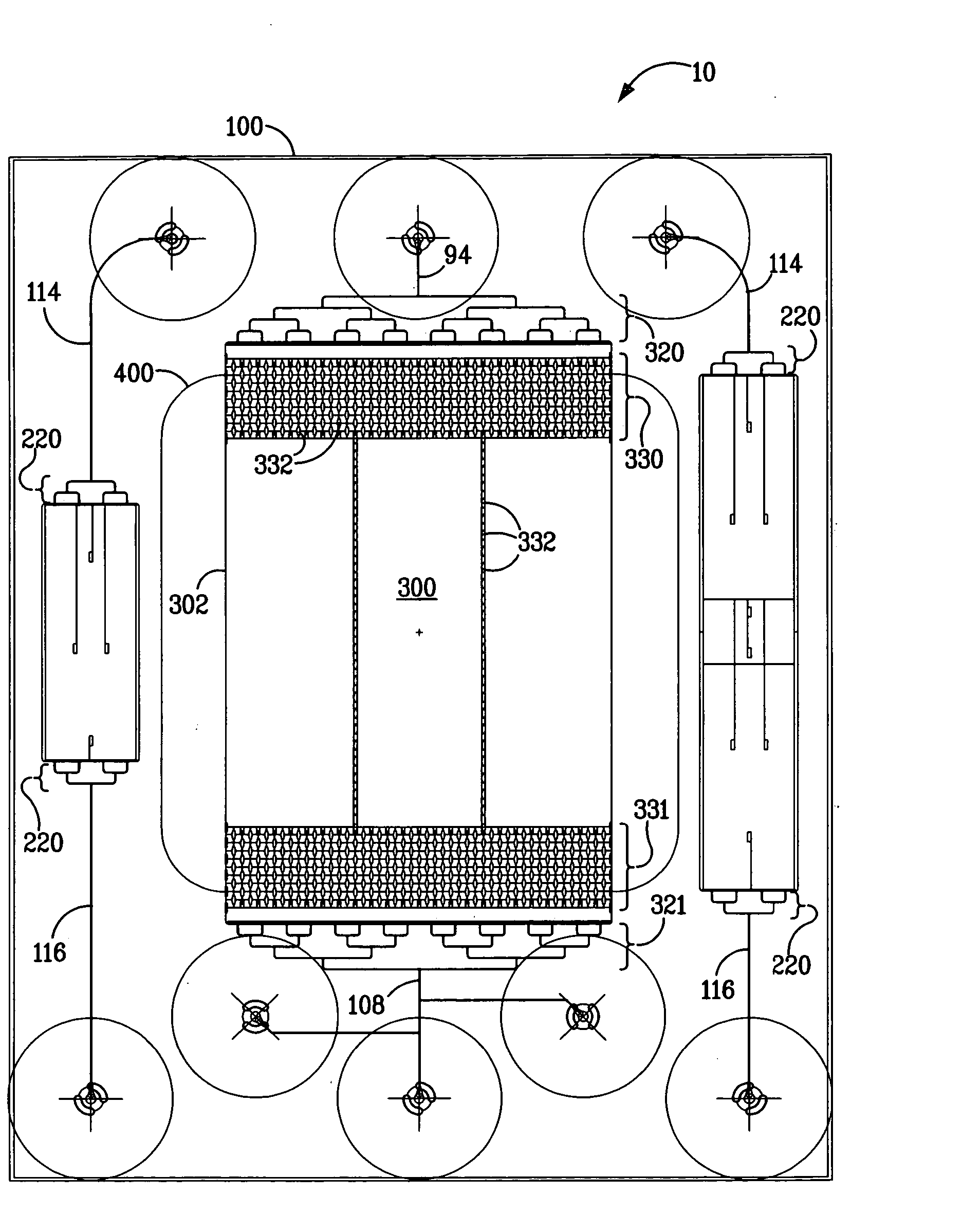
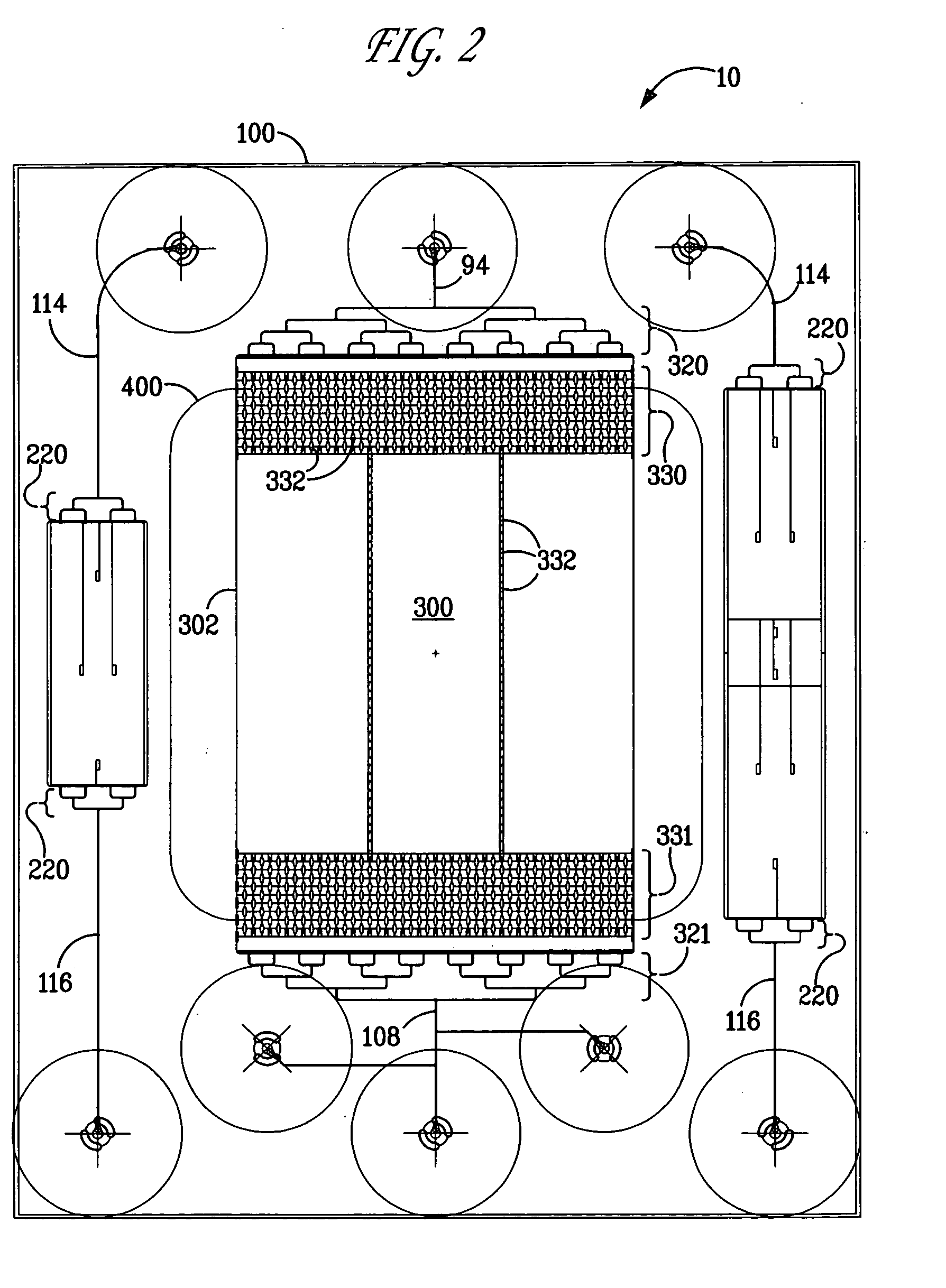
[0090] A microfluidic chip was fabricated containing a DNA gene microarray that is capable of detecting thousands of genes using a single experimental sample. The microfluidic chip of FIG. 8, inset, was produced in fused silica using standard wet-etching photolithography procedures. The chip layout is essentially the same as that depicted in FIG. 2. The compact design feature (2.5 cm×3.1 cm) enabled the production of six devices per wafer and enabled the use of low volumes of fluids suitable for gene microarray analysis. This microfluidic chip design also provided the ability to spot content genes after thermal bonding of the base and cover substrates (1100° C.). The open microarray probe design provided the ability to rapidly change the probe content sets. The open microarray design also enabled the performance of custom cDNA microarray construction (up to approximately 14000 spots) using a custom fabricated arrayer which can be programmed to deposit probes in complex architectures...


PUM

PropertyMeasurementUnit
thicknessaaaaaaaaaa
mean distanceaaaaaaaaaa
length scaleaaaaaaaaaa

Abstract

Disclosed are microflulidic chips that include a plurality of vias; a functionalized porous polymer monolith capable of being in fluid communication with a via; a microarray capable of being in fluid communication with the functionalized porous polymer monolith; and an observation port through which at least one target disposed within the microarray is capable of being detected. The disclosed microfluidic chips contain microarrays that can be effectively coupled to functionalized porous polymer monoliths for capturing and concentrating sample nucleic acids. Also disclosed are microfluidic chips containing microarray probes having observation ports that enable the preparation of microarrays and the detection of targets. These microfluidic chips are capable of capturing and concentrating genetic material for the analysis and identification of biological organisms, such as so-called “threat genes” from chimeric bioweapons.

Description

STATEMENT OF GOVERNMENT INTEREST [0001] This invention is made with Government support under contract no. DE-AC04-94AL85000 awarded by the U.S. Department of Energy to Sandia Corporation. The Government has certain rights in the invention.FIELD OF THE INVENTION [0002] The present invention is related to the field of microfluidic devices. The present invention is also related to the field of biological detection. BACKGROUND OF THE INVENTION [0003] Various scientific and patent publications are referred to herein. Each is incorporated by reference in its entirety. [0004] Recent advances in miniaturization have led to the development of microfluidic systems that are designed, in part, to perform a multitude of chemical and physical processes on a micro-scale. Typical applications include analytical and medical instrumentation, industrial process control equipment, liquid and gas phase chromatography, and the detection of biological weapons. In this context, there is a need for devices ...

Claims


Application Information

Patent Timeline
Patent Type & Authority Applications(United States)
IPC IPC(8): B01F5/06B01F13/00B01L3/00C12M1/34C12Q1/68
CPCB01F5/061B01L2400/086B01F15/0264B01F2005/0631B01J2219/00286B01J2219/00432B01J2219/00511B01J2219/00576B01J2219/00605B01J2219/0061B01J2219/00612B01J2219/00626B01J2219/00637B01J2219/00641B01J2219/00657B01J2219/00659B01J2219/00664B01J2219/00691B01J2219/00722B01J2219/00729B01L3/5023B01L3/5027B01L2300/0636B01L2300/0681B01L2300/0816B01L2300/0867B01L2300/087B01L2300/0877B01L2300/0883B01L2400/0418B01L2400/0421B01L2400/0487B01F13/0059B01F25/4319B01F33/30B01F35/7182
Inventor WEST, JASON A.A.SHEPODD, TIMOTHY J.GRIFFITHS, STEWART K.RENZI, RONALD F.WIEDENMAN, BOYD J.
Owner SANDIA NAT LAB
Learn More