
Th detecting method for non-heparin anticoagulant blood sample

A blood sample and detection method technology, applied in the field of Th detection of non-heparin anticoagulated blood samples, can solve the problems of sample type limitation, re-collection, sample waste, etc., and achieve the effect of avoiding repeated collection, avoiding sample waste, and expanding the scope

Active Publication Date: 2020-01-10
杭州联科生物技术股份有限公司
View PDF6 Cites 0 Cited by
  • Summary
  • Abstract
  • Description
  • Claims
  • Application Information

AI Technical Summary

Problems solved by technology

[0006] Aiming at the deficiencies of the prior art, the present invention provides a method for Th detection of non-heparin anticoagulated blood samples, aiming to solve the problem that non-heparin anticoagulated blood samples will affect the activation of Th cells and the secretion of cytokines during Th detection. Cytokines cannot be detected, which limits the types of samples that can be tested, resulting in the problem of sample waste or re-collection

Method used


Image

  • Th detecting method for non-heparin anticoagulant blood sample

Examples


Embodiment 1

[0031] A Th detection method of a non-heparin anticoagulated blood sample, comprising the following steps:

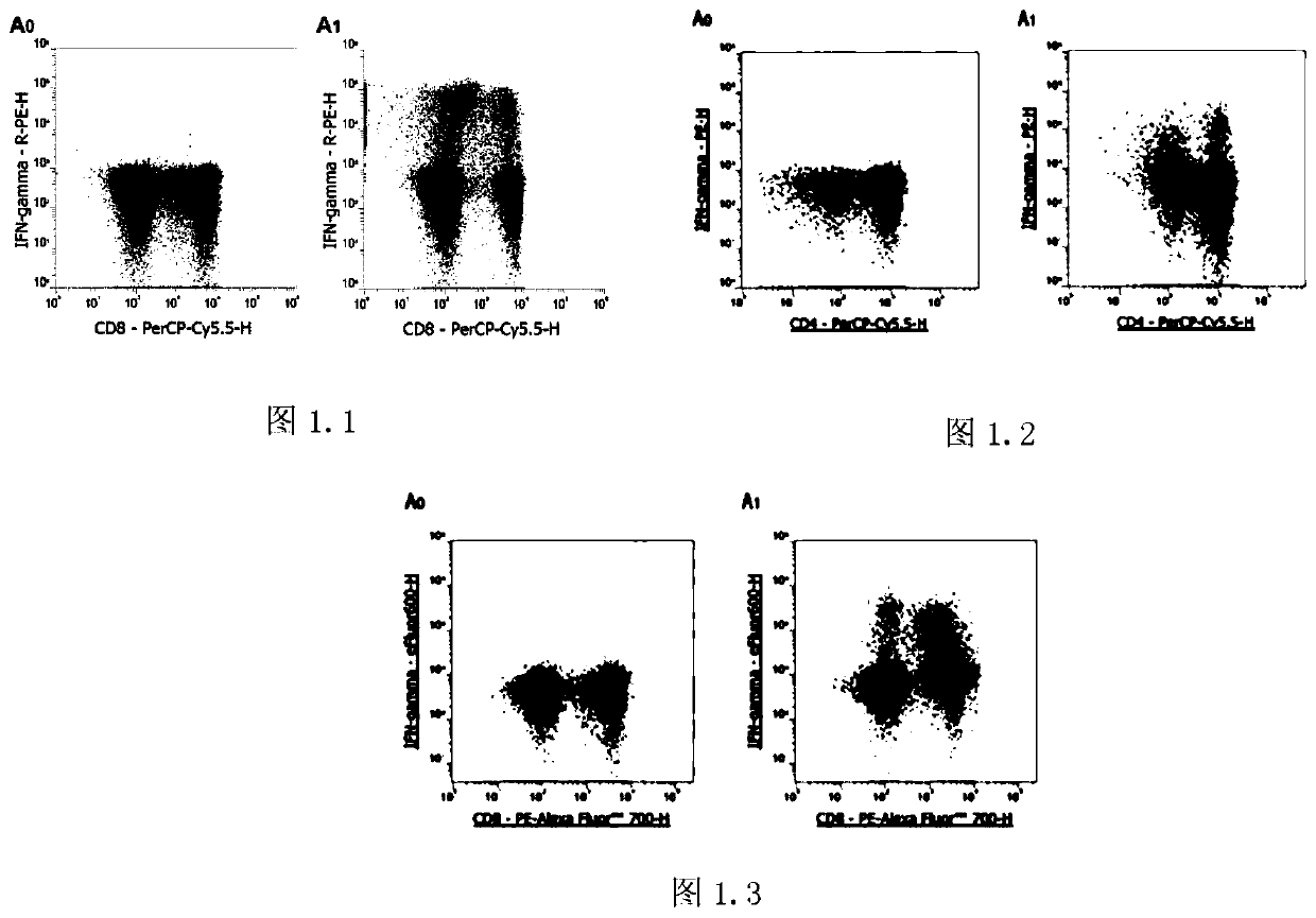
[0032] Step 1: Separate peripheral blood mononuclear cells (PBMCs) with lymphocyte separation medium, resuspend the pellet with medium containing 10% fetal bovine serum, and make the cell concentration 1×10 7 / ml, take 250μl PBMCs to the flow tube, add 1μl PMA / Ionomycin Mixture (250×) and 1μl BFA / Monensin Mixture (250×), use only PBMCs as a control, mix well, and incubate at 37℃ for 4- 6 hours, take out every 1-2 hours and mix well;

[0033] Step 2: Take 100 μl of cell suspension from the sample tube and the control tube into a new flow tube, add the corresponding flow antibody, shake and mix, and incubate at room temperature in the dark for 15 minutes;

[0034] Step 3: Add 100 μl FIX&PERM Medium A to each tube, vortex to mix, and incubate at room temperature in the dark for 15 minutes;

[0035] Step 4: Dilute 10×Flow Cytometry Staining Buffer to 1× with distilled wat...


PUM


Abstract

The invention discloses a Th detecting method for a non-heparin anticoagulant blood sample. In the method, a non-heparin anticoagulant sample is processed by using a lymphocyte separation solution toobtain peripheral blood mononuclear cells (PBMC), the effect of the anticoagulant on cell activation and cytokine secretion is removed, and the serum is added at the same time in the stimulation / blocking stage to provide cells with sufficient nutrients. After antibody staining and fixed membrane rupture, the method can be successfully used for the detection of Th cytokines, thereby expanding the range of Th detection sample type, enables researchers or testers to make full use of samples for Th detection, avoids waste and repeated collection of samples caused by the collection of non-heparin anticoagulant samples, and greatly improves efficiency.

Description

technical field [0001] The invention relates to a Th detection method of a non-heparin anticoagulated blood sample. Background technique [0002] Commonly referred to as Th1, Th2 and Th17 refer to T helper cells capable of differentiating into Th1, Th2 and Th17 under various physiological and pathological conditions (Th0 cells in the strict sense). In a resting state (that is, without any stimulation, such as the normal physiological state of a person), the ability of Th0 to differentiate into Th1, Th2 and Th17 is very weak, so the peripheral blood only contains a very small amount of Th1, Th2 and Th17 cells. IFN-γ, IL-4 and IL-17A were also minimally detectable. When Th cells are stimulated by external factors (such as stimulants, pathogens, etc.), Th0 will differentiate into Th1, Th2 or Th17, and the specific differentiation trend depends on the type of cytokines. At this time, more IFN-γ, IL-4 or IL-17A was also detected. The Th1, Th2 and Th17 detected in the experimen...

Claims


Application Information

Patent Timeline
IPC IPC(8): G01N15/14G01N1/28G01N1/30
CPCG01N15/14G01N1/28G01N1/30G01N2015/1006G01N15/01
Inventor 黄振宇
Owner 杭州联科生物技术股份有限公司
Learn More