Method for estimating tumor pose in closed phantom

A pose estimation and closed body technology, applied in the field of breath tracking system, can solve the problems of complex operation and high cost, and achieve the effect of convenient operation, simple structure and accurate positioning

Active Publication Date: 2020-01-10
SUZHOU UNIV
View PDF4 Cites 0 Cited by
  • Summary
  • Abstract
  • Description
  • Claims
  • Application Information

AI Technical Summary

Problems solved by technology

The main way to obtain the movement of the tumor in the phantom is to implant markers into the simulated tumor, and obtain the movement of the simulated tumor with the help of imaging technology such as X-ray, but the cost is high and the operation is relatively complicated

Method used


Image

  • Method for estimating tumor pose in closed phantom
  • Method for estimating tumor pose in closed phantom
  • Method for estimating tumor pose in closed phantom

Examples


Embodiment Construction

[0037] The present invention will be further described below in conjunction with the accompanying drawings and specific embodiments, so that those skilled in the art can better understand the present invention and implement it, but the examples given are not intended to limit the present invention.

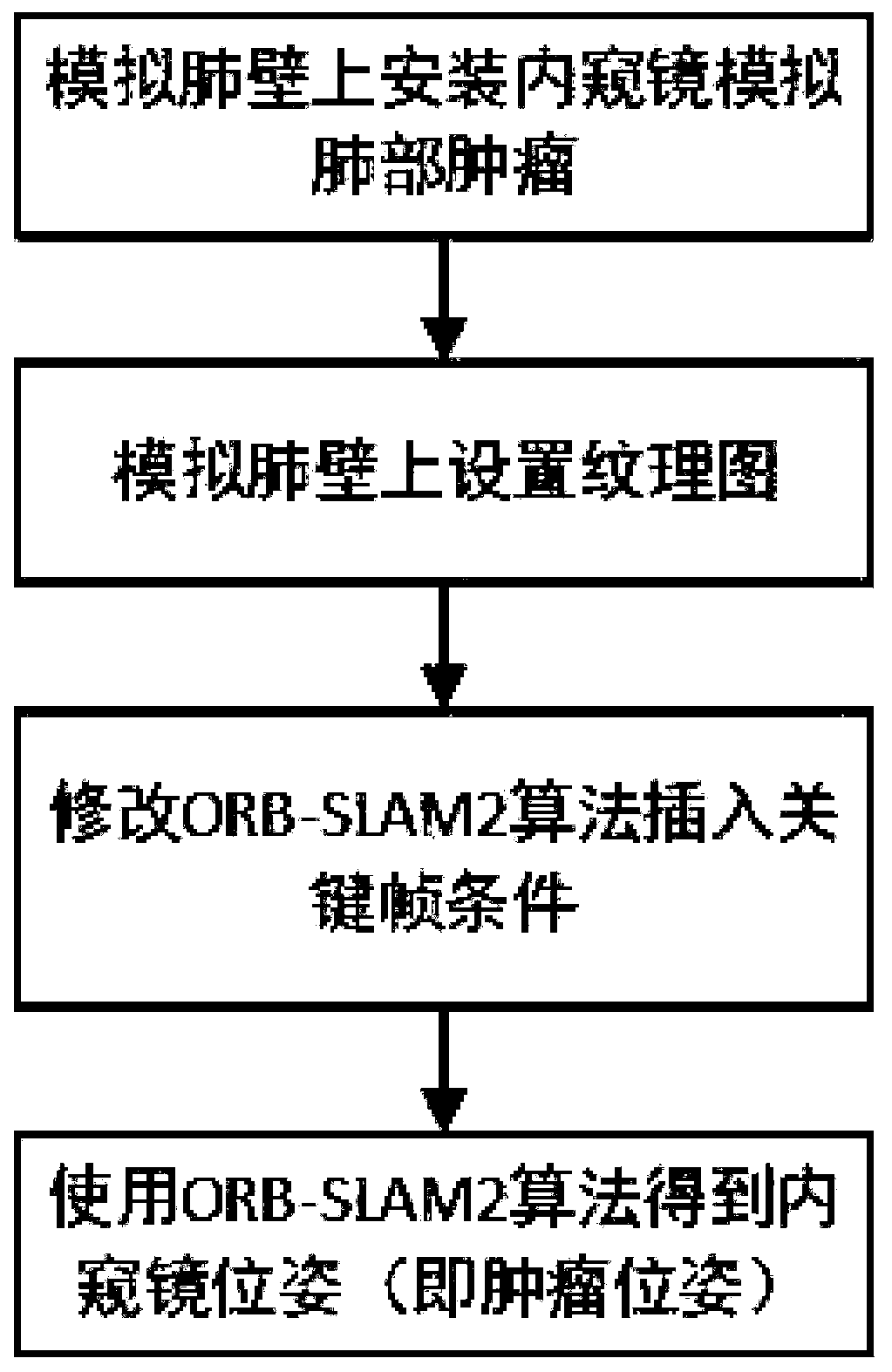
[0038] refer to figure 1 As shown, the present invention discloses a method for estimating the pose of a tumor in a closed phantom, comprising the following steps:

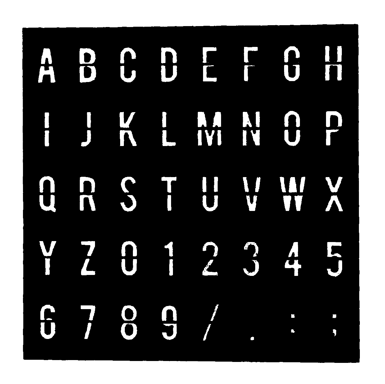
[0039] Step 1: Fix and set the visual texture map on the inner wall of the simulated lung. Since the simulated lung will deform when simulating breathing movement, in order to avoid the visual texture map from moving, the visual texture map needs to be fixed. The specific operation is to cut a hole in the simulated lung first, and paste the hard board of the texture map inside the lung. A hard board of the same size is pasted on the corresponding position outside the lung for reinforcement. image 3 is a visual tex...


PUM


Abstract

The invention discloses a method for estimating a tumor pose in a closed phantom. The method comprises the following steps of fixedly setting a visual texture map on the inner wall of a simulated lung; installing a monocular endoscope on the wall surface of the simulated lung to simulate a lung tumor, wherein the monocular endoscope is arranged towards the visual texture map; when the simulated lung moves, using the monocular endoscope to collect a video frame of the visual texture map; processing the video frame by using an ORB-SLAM2 algorithm, re-establishing a key frame discrimination criterion, obtaining the key frame, and solving the pose of the monocular endoscope; and obtaining the simulated tumor pose data according to the pose of the monocular endoscope. According to the present invention, the tumor pose in the closed phantom is estimated, the structure is simple, the operation is convenient, and the positioning is accurate.

Description

technical field [0001] The invention relates to the technical field of a breathing tracking system of a radiotherapy robot, in particular to a method for estimating the pose of a tumor in a closed phantom. Background technique [0002] Tumors in the human thoracoabdominal cavity, especially lung tumors, are usually accompanied by breathing movements. The precise positioning of tumors in motion is an important part of the research on radiotherapy robots today. Since the tumor is located inside the human body, for the acquisition of the motion of the internal tumor, clinically, gold labels are mostly implanted in or around the tumor, and X-ray imaging equipment is used to obtain the position coordinates of the tumor target area. Since long-term exposure to X-rays will cause damage to normal tissues around the tumor, it is not advisable to use imaging techniques such as X-rays to directly locate the tumor in real time. [0003] For this reason, using body surface information ...

Claims


Application Information

Patent Timeline
Patent Type & Authority Applications(China)
IPC IPC(8): G06T7/40G06T7/73G06T17/00G06K9/00
CPCG06T17/00G06T7/40G06T7/73G06T2207/30061G06T2207/30096G06V20/46
Inventor 郁树梅后鹏程孙荣川匡绍龙孙立宁
Owner SUZHOU UNIV
Why Eureka
Try Eureka