Abdomen multi-organ nuclear magnetic resonance image segmentation method and system based on FCN and medium

A nuclear magnetic resonance image, multi-organ technology, applied in the direction of instruments, computer components, biological neural network models, etc., can solve the problem of lack of transformation of low-level features, and achieve high segmentation accuracy and convenient and fast operation

Active Publication Date: 2020-01-17
SUN YAT SEN UNIV
View PDF3 Cites 21 Cited by
  • Summary
  • Abstract
  • Description
  • Claims
  • Application Information

AI Technical Summary

Problems solved by technology

[0006] The U-Net network structure uses the skip connection method to directly fuse the low-level feature map with the high-level feature map after upsampling (deconvolution),

Method used


Image

  • Abdomen multi-organ nuclear magnetic resonance image segmentation method and system based on FCN and medium
  • Abdomen multi-organ nuclear magnetic resonance image segmentation method and system based on FCN and medium
  • Abdomen multi-organ nuclear magnetic resonance image segmentation method and system based on FCN and medium

Examples


Embodiment Construction

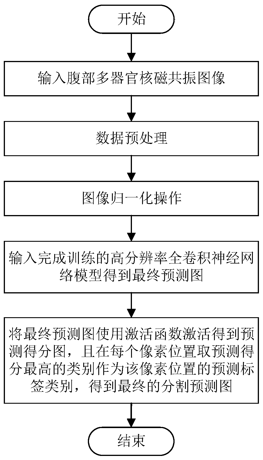
[0041] In the following, we will take the segmentation of abdominal multi-organ MR images as an example in five different regions: no organ region (C0), liver region (C1), right kidney region (C2), left kidney region (C3) and spleen region (C4) , the method, system and medium of the FCN-based abdominal multi-organ nuclear magnetic resonance image segmentation of the present invention will be further described in detail.

[0042] Such as figure 1 As shown, the implementation steps of the FCN-based abdominal multi-organ MRI image segmentation method in this embodiment include:

[0043] 1) Obtain the input abdominal multi-organ MRI image and perform data preprocessing and image normalization operations;

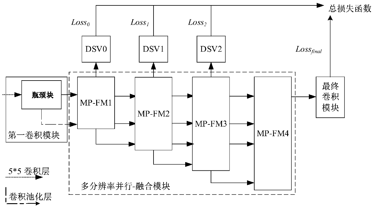
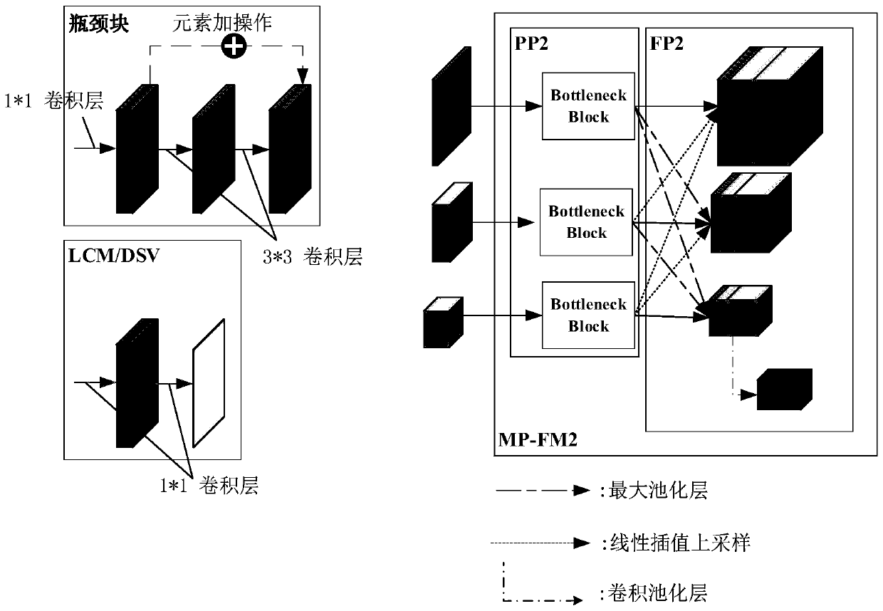
[0044] 2) Input the normalized multi-organ MRI images of the abdomen into the trained high-resolution fully convolutional neural network model to obtain the final prediction map. The high-resolution fully convolutional neural network model has been pre-trained to establish a no...


PUM


Abstract

The invention discloses an abdominal multi-organ nuclear magnetic resonance image segmentation method and system based on FCN, and a medium. The abdominal multi-organ nuclear magnetic resonance imagesegmentation method comprises the following implementation steps: acquiring an input image and carrying out data preprocessing and image normalization operation; inputting the normalized abdominal multi-organ nuclear magnetic resonance image into a trained high-resolution full convolutional neural network model to obtain a final prediction image, wherein the high-resolution full convolutional neural network model is pre-trained to establish a mapping relationship between the normalized abdominal multi-organ nuclear magnetic resonance image and the corresponding final prediction image; and activating the final prediction graph by using an activation function to obtain a prediction score graph, and taking a category with the highest prediction score at each pixel position as a prediction label category of the pixel position to obtain a final segmentation prediction graph. According to the abdominal multi-organ nuclear magnetic resonance image segmentation method, automatic segmentation of the abdominal multi-organ nuclear magnetic resonance image can be realized, for example, the abdominal multi-organ MR image is segmented according to five different region types of an organ-free region, a liver region, a right kidney region, a left kidney region and a spleen region.

Description

technical field [0001] The invention relates to the fields of digital medical image processing and analysis and computer-aided diagnosis, and in particular to an FCN-based method, system and medium for segmenting abdominal multi-organ nuclear magnetic resonance images. Background technique [0002] Understanding the prerequisites for complex medical procedures plays an important role in the success of the surgery. To enrich the level of understanding, physicians use advanced tools such as 3D visualization and printing, which are required to extract objects of interest from DICOM images. Accurate segmentation of multiple abdominal organs (i.e., liver, kidney, and spleen) is critical for several clinical procedures, including but not limited to liver pre-evaluation for living donor-based transplantation or detailed analysis of multiple abdominal Prior to arterial surgery, in order to properly position the vessels of the graft and access them. This prompts the ongoing researc...

Claims


Application Information

Patent Timeline
IPC IPC(8): G06K9/34G06N3/04
CPCG06V10/267G06V2201/03G06N3/045
Inventor 戈峰肖侬卢宇彤陈志广邓楚富
Owner SUN YAT SEN UNIV
Why Eureka
Try Eureka