Three-dimensional reconstruction method and device for blood vessel image

A blood vessel image and three-dimensional reconstruction technology, applied in the field of medical image processing, can solve the problem that two-dimensional tomographic images cannot segment pulmonary blood vessels, and achieve the effect of reducing damage and improving accuracy.

Active Publication Date: 2020-01-17
西人马大周(深圳)医疗科技有限公司
View PDF7 Cites 5 Cited by
  • Summary
  • Abstract
  • Description
  • Claims
  • Application Information

AI Technical Summary

Problems solved by technology

[0004] The embodiment of the present invention provides a method and device for three-dimensional reconstruction of blood vessel images, which solves the problem that pulmonary blood vessels cannot be segmented from two-dimensional tomographic images

Method used


Image

  • Three-dimensional reconstruction method and device for blood vessel image
  • Three-dimensional reconstruction method and device for blood vessel image
  • Three-dimensional reconstruction method and device for blood vessel image

Examples


Embodiment Construction

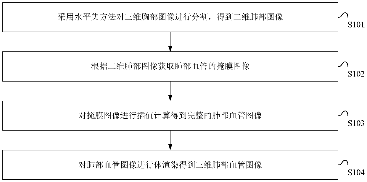
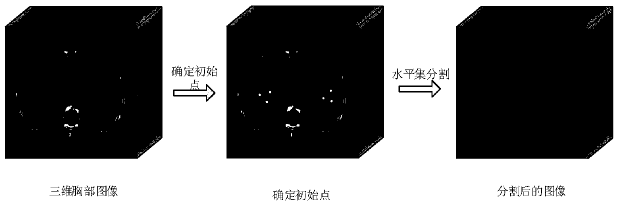
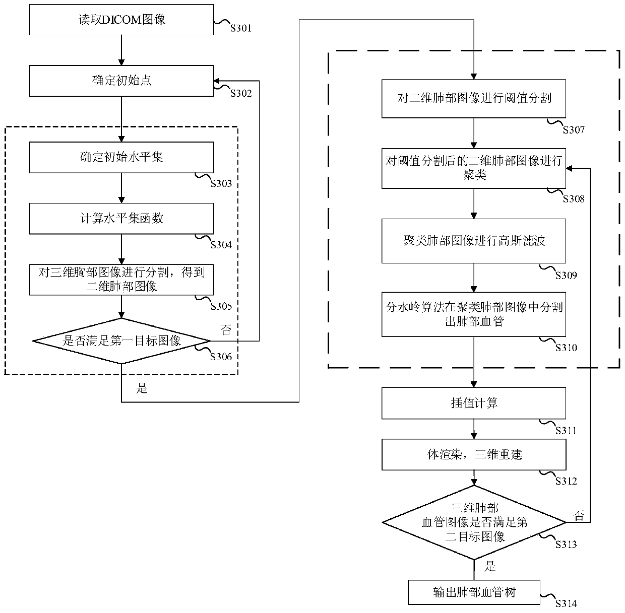
[0058] The characteristics and exemplary embodiments of various aspects of the present invention will be described in detail below. In order to make the purpose, technical solutions and advantages of the present invention more clear, the present invention will be further described in detail below in conjunction with the accompanying drawings and embodiments. It should be understood that the specific embodiments described here are only configured to explain the present invention, not to limit the present invention. It will be apparent to one skilled in the art that the present invention may be practiced without some of these specific details. The following description of the embodiments is only to provide a better understanding of the present invention by showing examples of the present invention.

[0059] It should be noted that in this article, relational terms such as first and second are only used to distinguish one entity or operation from another entity or operation, and ...


PUM


Abstract

The embodiment of the invention provides a three-dimensional reconstruction method and device for a blood vessel image. The method comprises the following steps: segmenting a three-dimensional chest image by adopting a level set method to obtain a two-dimensional lung image; obtaining a mask image of a lung blood vessel according to the two-dimensional lung image; performing interpolation calculation on the mask image to obtain a complete lung blood vessel image; and performing volume rendering on the lung blood vessel image to obtain a three-dimensional lung blood vessel image. According to the embodiment of the invention, a pulmonary vessel tree can be established to assist a doctor in avoiding vessels as much as possible when determining an operation scheme, so that damage to the vessels is reduced.

Description

technical field [0001] The invention relates to the technical field of medical image processing, in particular to a method and device for three-dimensional reconstruction of blood vessel images. Background technique [0002] Affected by factors such as air pollution and smoking, lung diseases have seriously endangered human health and life. When planning lung surgery clinically, doctors hope to observe the shape and spatial position of some important tissues such as blood vessels, diseased bodies, and sensitive tissues in the lungs, and then determine a scientific surgical plan. [0003] At present, the clinical diagnosis of the lungs mostly uses two-dimensional tomographic images, but the two-dimensional tomographic images cannot provide an intuitive lung structure for medical diagnosis, especially the pulmonary blood vessels are small in size and low in contrast compared with other lung tissues, and The bronchi interlaced with the lungs, and the structure is complex. Ther...

Claims


Application Information

Patent Timeline
Patent Type & Authority Applications(China)
IPC IPC(8): G06T7/11G06T7/136G06T7/155G06T17/00
CPCG06T7/11G06T7/136G06T7/155G06T17/00G06T2210/41G06T2207/20152G06T2207/30061G06T2207/30101
Inventor 聂泳忠赵银妹吕明涛赵春莲
Owner 西人马大周(深圳)医疗科技有限公司
Why Eureka
Try Eureka