A kind of preparation method of lysosomal membrane coating nanoparticle

A nanoparticle and lysosomal membrane technology, which is applied to medical preparations with non-active ingredients, medical preparations containing active ingredients, and pharmaceutical formulas, etc., can solve the damage, loss and low encapsulation efficiency of biologically active proteins in membrane structures. And other issues

Active Publication Date: 2020-10-27
BEIJING UNIV OF CHEM TECH
View PDF12 Cites 0 Cited by
  • Summary
  • Abstract
  • Description
  • Claims
  • Application Information

AI Technical Summary

Problems solved by technology

These passive coating methods have low loading efficiency and are easy to cause damage and loss to the membrane structure and biologically active proteins

Method used


Image

  • A kind of preparation method of lysosomal membrane coating nanoparticle
  • A kind of preparation method of lysosomal membrane coating nanoparticle
  • A kind of preparation method of lysosomal membrane coating nanoparticle

Examples


Embodiment 1

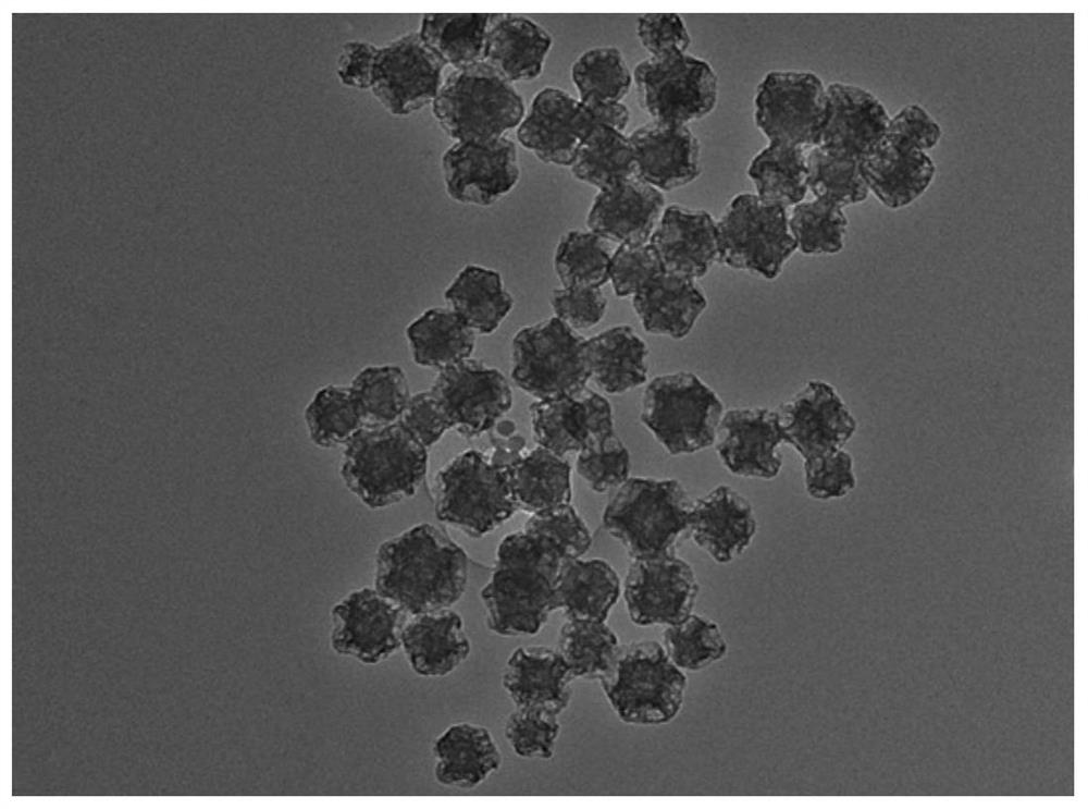
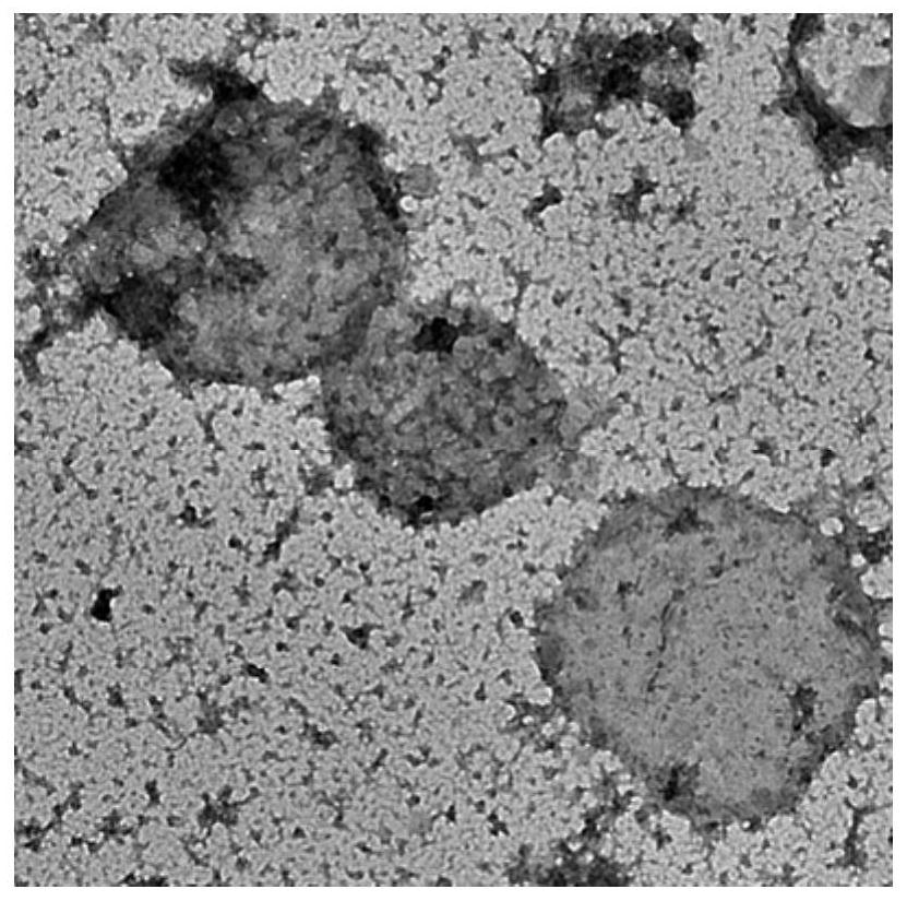
[0048] Nanoparticle PMCS TEM figure 1 As shown, the size of PMCS nanoparticles is 140 nm. TEM image of lysosome figure 2 As shown, the lysosome size is 450 nm.

example 2

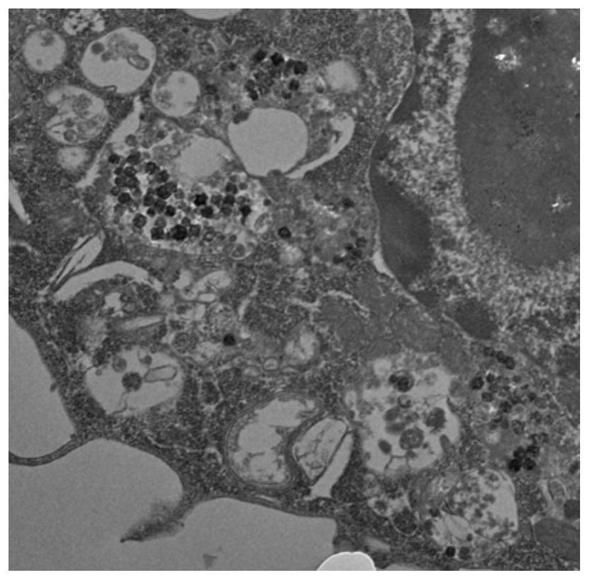
[0050] Observation of macrophages engulfing PMCS nanoparticles and internalizing them in lysosomes. First, macrophages were inoculated into 6-well plates at a concentration of 400,000 cells per well, and incubated in an incubator. Replace the old culture medium in the 6-well plate with 50 μg / mL PMCS nanoparticles dispersed in DMEM. Cells were washed with PBS buffer and collected by centrifugation. Wash twice with PBS. The pellet was fixed in a PBS solution with a volume fraction of 2.5% glutaraldehyde and 4% paraformaldehyde. biological transmission electron microscope Figure 4 As shown, the extracted lysosomal membrane-coated nanoparticle PMCS is a vesicle-like particle with a single-layer membrane structure on the outside, with a particle size of 450 nm, and contains PMCS nanoparticles inside. In addition, the three potentials of PMCS, lysosome and lysosomal membrane-coated nanoparticle PMCS are as follows: Figure 5 shown. They are: -5.6mV, -24.7mV, -23.2mV.

Embodiment 3

[0052] (1) Macrophages were cultured in DMEM containing 10% fetal calf serum, and 100 μL of cell suspension was prepared in a 96-well plate, and 5000 cells were plated per well. The plates were pre-incubated in the incubator. After the cells adhered to the wall, the activated macrophages were stimulated for 1 h with 100 μL of lipopolysaccharide at a final concentration of 10 μg / mL. Then remove the culture medium.

[0053] (2) Add 100 μL of nanoparticles PMCS with different concentrations (6.25, 12.5, 25, 50, 100, 200 μg / mL) to the culture plate.

[0054] (3) Continue to incubate for 24 hours, add 100 μL of CCK-8 solution to each well, incubate the culture plate in the incubator for 1.5 hours, measure the absorbance with a microplate reader, and process the data. Such as Image 6 As shown, when the concentration of PMCS is greater than 50 μg / mL, the incubation time is 24 hours, and the activity of macrophages is lower than 60%. Under the premise of ensuring the activity of m...


PUM

PropertyMeasurementUnit
sizeaaaaaaaaaa
sizeaaaaaaaaaa
particle diameteraaaaaaaaaa

Abstract

The invention relates to a preparation method for a lysosomal membrane coated nanoparticle. Under the function of the driving of macrophages, an active lysosomal membrane coated nanoparticle is synthesized. Specifically, the preparation method comprises the following steps of: firstly, activating the macrophages by lipopolysaccharides, stimulating the secretion of lysosomal related proteases in the macrophages, and enhancing the enzymatic activity of the lysosomal related proteases; then, utilizing the phagocytic ability of the macrophages to jointly incubate the prepared nanoparticle and themacrophages to enable the macrophages to carry out phagocytosis on the nanoparticle, and carrying out internalization on the nanoparticle in macrophage lysosome; and finally, utilizing a lysosome extraction kit to extract the lysosome to obtain the lysosomal membrane coated nanoparticle. Compared with a preparation method for a traditional lipid membrane coated nanoparticle, on one hand, the preparation method disclosed by the invention is simple, and does not need to consider damage caused for the membrane by pressure or electric shock. On the other hand, the macrophages are taken as drivingforce, and the prepared lysosomal membrane coated nanoparticle keeps the activity of hydrolase in the lysosome.

Description

Technical field: [0001] The invention relates to a preparation technology and method of lysosome-coated functional nano-medicines, which synthesize active lysosome membrane-coated nanoparticles under the action of macrophage drive. Specifically, firstly, lipopolysaccharide is used to activate macrophages, stimulate the secretion of lysosome-associated proteases in macrophages and enhance their enzymatic activity; , making macrophages phagocytose nanoparticles, internalizing nanoparticles in macrophage lysosomes, and finally using a lysosome extraction kit to extract lysosomes to obtain nanoparticles coated with lysosome membranes. Background technique: [0002] Due to the diversity, heterogeneity and recurrence of tumors, it is difficult to effectively inhibit tumor recurrence and metastasis by relying solely on traditional surgery, radiotherapy and chemotherapy. For a long time, improving the targeting and efficacy of drugs has been the direction of continuous efforts of r...

Claims


Application Information

Patent Timeline
Patent Type & Authority Patents(China)
IPC IPC(8): A61K47/46A61K9/51A61K38/46A61K41/00A61P35/00
CPCA61K9/5176A61K9/5192A61K38/46A61K41/0033A61P35/00A61K2300/00
Inventor 刘惠玉魏炜李闪闪
Owner BEIJING UNIV OF CHEM TECH
Why Eureka
Try Eureka