Pathological image processing method and system

A technology of pathological images and processing methods, applied in the field of image processing, to achieve the effect of fast and accurate diagnosis, convenience and no need for scanning procedures

Pending Publication Date: 2020-02-07
杭州迪英加科技有限公司
View PDF10 Cites 8 Cited by
  • Summary
  • Abstract
  • Description
  • Claims
  • Application Information

AI Technical Summary

Problems solved by technology

[0004] One purpose of the present invention is to solve the problem in the prior art that the cell smear needs to be scanned into a full-field image under a high-magnification microscope, and then uploaded to a diagnostic tool for analysis. The present invention provides a pathological image processing method and system

Method used


Image

  • Pathological image processing method and system
  • Pathological image processing method and system
  • Pathological image processing method and system

Examples


Embodiment Construction

[0031] The present invention will be further described below in conjunction with accompanying drawing:

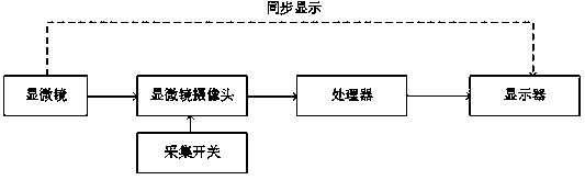
[0032] The microscope used in this embodiment is an in-line microscope, including 1 eyepiece and 4 objective lenses; the magnification of the eyepiece is 10 times, the magnification of the objective lens is 4 times, 10 times, 20 times, 40 times, and the microscope camera The MPP values ​​of the collected images are 1.5, 0.6, 0.3 and 0.15, respectively.

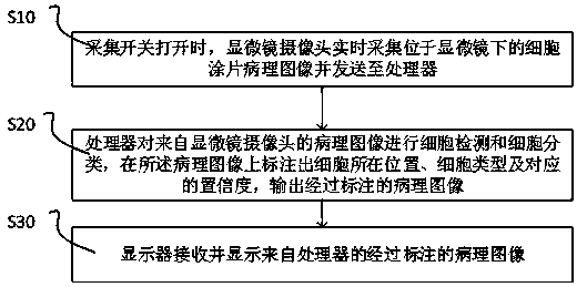
[0033] A pathological image processing system, such as figure 1As shown, including: microscope, microscope camera, acquisition switch, processor and display. The microscope is used to observe the cell smear; the microscope camera is connected with the microscope lens and used to collect pathological images of the cell smear under the microscope. The pathological images collected by the microscope camera can be PNG, JPG, mrxs, svs, kfb, ndpi, etc., and can be video data or picture data. If it is video data, because there a...


PUM


Abstract

The invention relates to a pathological image processing system. The pathological image processing system comprises a microscope, a microscope camera, an acquisition switch, a processor and a display,wherein the microscope is used for observing the cell smear; the microscope camera is connected with the microscope lens and is used for collecting pathological images of the cell smear under the microscope; the processor is in communication connection with the microscope camera, receives a pathological image from a microscope camera, preprocesses the pathological image, carries out cell detection and cell classification on the processed pathological image, marks a cell position, a cell type and a corresponding confidence coefficient on the pathological image, and outputs the marked pathological image; the display is connected with the processor and is used for receiving and displaying the marked pathological image; and the acquisition switch is arranged on the microscope camera and controls the operation of the microscope camera.

Description

technical field [0001] The invention relates to the field of image processing, in particular to a pathological image processing method and system. Background technique [0002] Cell pathology (cytopathology), based on histopathology, is a discipline that elucidates the nature of diseases by carefully observing cell morphology, cell arrangement, and intercellular relationships. It is an important part of pathology. The cell sample is made into a cell smear through sampling, fixing, preparation, staining and other processes. The cytopathologist observes the cell components in the cell smear under a microscope for diagnosis, first browses the whole under the low-power objective lens, and turns to Carefully observe the target cells under a high-power objective lens. [0003] The currently used computer-aided reading systems include: ThinPrep Imaging System (TIS) and BDFocalPoint reading device, both of which are microscope-based slide scanning analysis systems, which improve th...

Claims


Application Information

Patent Timeline
IPC IPC(8): G06K9/00G06K9/62G06N3/08
CPCG06N3/08G06V20/69G06V20/693G06F18/253G06F18/214
Inventor 韩鑫李涵生宋敏敏杨林崔磊
Owner 杭州迪英加科技有限公司
Why Eureka
Try Eureka