Pulmonary nodule morphological classification method based on neural network

A technology of morphological classification and neural network, applied in biological neural network model, neural architecture, image analysis, etc., can solve the problems of limited effect of 3D CT images, loss of size information, time-consuming and labor-intensive, etc., to save manpower and material resources, Quickly detect and realize the effect of batch operations

Inactive Publication Date: 2020-02-07
WEST CHINA HOSPITAL SICHUAN UNIV +2
View PDF7 Cites 23 Cited by
  • Summary
  • Abstract
  • Description
  • Claims
  • Application Information

AI Technical Summary

Problems solved by technology

Compared with traditional machine learning methods, the operation is complex, time-consuming and labor-intensive - the accuracy of classification depends entirely on the extraction of features, that is, the definition of feature calculation and the selection of feature sets; if segmentation is required before feature extraction , the expression effect of its feature set is also directly related to the accuracy of the segmentation method, and for CT images with many blood vessels overlapping with pulmonary nodules and other lung tissues, this task is extremely challenging; except In addition, this type of method can only extract shallow features, and its effect in complex 3D CT images is limited.
However, for some current neural network nodule classification methods, they regard the 3D CT lung image as multiple 2D slices, directly intercept the nodules to be classified from the whole slice as data, and use an independent data in the data set for binary The training of three-dimensional network model, this method obviously cannot extract the features shown in the three-dimensional space of nodules; in addition, due to the growth of nodules, its diameter is an important factor affecting the morphological characteristics and properties, the previous method adopts Using a fixed-size image as the input of the network, the key size information of the nodule will be lost to a certain extent or even completely.

Method used


Image

  • Pulmonary nodule morphological classification method based on neural network
  • Pulmonary nodule morphological classification method based on neural network
  • Pulmonary nodule morphological classification method based on neural network

Examples


Embodiment Construction

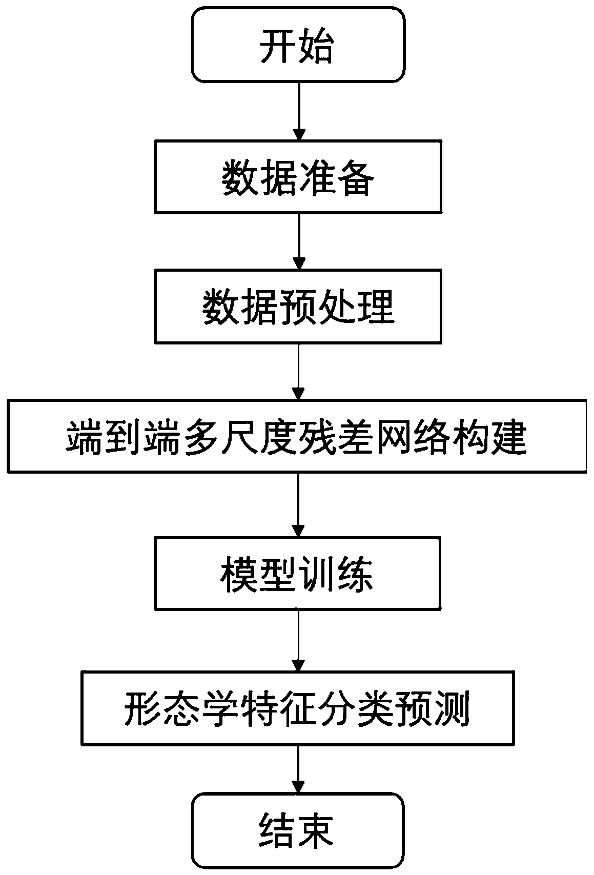
[0056] In order to make the object, technical solution and advantages of the present invention clearer, the present invention will be further described in detail below in conjunction with the accompanying drawings.

[0057] The present invention comprises the following steps:

[0058] 1. Data preparation. This stage mainly completes the data import from the hospital data system and the calibration of the pulmonary nodules to be classified.

[0059] 2. Data preprocessing. This stage preprocesses the data, mainly including the preprocessing of the original CT image and the interception of the nodule area.

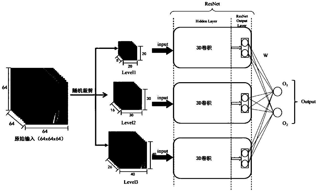
[0060] 3. Construct a multi-scale 3D residual network model, and use the collected data and calibration results to train the model.

[0061] 4. Repeat step 3 to build and train models for various morphological classifications.

[0062] 5. Use the model trained in step 3 and step 4 to classify the lung nodules automatically detected by the detection task, and output the in...


PUM


Abstract

The invention discloses a pulmonary nodule morphological classification method based on a neural network. The pulmonary nodule morphological classification method comprises the following steps of 1, data preparation; wherein in the stage, data is imported from a hospital data system, and pulmonary nodules to be classified are calibrated; 2, data preprocessing: preprocessing the data at the stage,mainly including preprocessing of an original CT image and interception of a nodule region; 3, constructing a multi-scale three-dimensional residual network model, and training the model by using theacquired data and a calibration result; 4, repeating the step 3, and constructing and training various morphological classification models; and 5, classifying the pulmonary nodules automatically detected by the detection task by using the models trained in the steps 3 and 4, and outputting pulmonary nodule morphological characteristic information predicted by the model. According to the method, three scales of nodules are intercepted to serve as input of a network of corresponding scale residuals, depth features of the nodules of the three scales are extracted, and finally a final classification result is obtained through fusion and integration of three models.

Description

technical field [0001] The invention relates to the technical field of deep learning, in particular to a neural network-based morphological classification method for pulmonary nodules. Background technique [0002] CAD systems for lung cancer have been extensively studied in recent years and mainly include two tasks: lung nodule detection and classification. Judging the nature of nodules through morphological features in the classification task of nodules is also receiving continuous attention. Jeremy J. Erasmus, John E. Connolly, H. Page McAdams and Victor L. Roggli in their early From a medical point of view, the study pointed out the reliability of judging the properties of nodules by their morphological characteristics, and proved the relationship between the characteristics of nodules and their properties through actual data. Several important features related to the nature are also noted, including spiculation, lobulation, texture, and calcification. [0003] Many ex...

Claims


Application Information

Patent Timeline
Patent Type & Authority Applications(China)
IPC IPC(8): G06K9/62G06N3/04G06T7/00
CPCG06T7/0012G06T2207/10081G06T2207/20081G06T2207/20084G06T2207/30064G06N3/045G06F18/241
Inventor 白红利章毅王成弟郭际香李为民徐修远邵俊易乐甘云翠赵科甫陈思行周凯
Owner WEST CHINA HOSPITAL SICHUAN UNIV
Why Eureka
Try Eureka