Unconstrained scanning and voxel-based three-dimensional real-time bone imaging method

An imaging method and unconstrained technology, applied in the fields of medical imaging and ultrasonic bone imaging, which can solve the problems of uneven scanning speed, changes in scanning range, and different scanning methods and attitudes.

Active Publication Date: 2020-04-07
SHANGHAI TECH UNIV
View PDF3 Cites 2 Cited by
  • Summary
  • Abstract
  • Description
  • Claims
  • Application Information

AI Technical Summary

Problems solved by technology

In addition, limited by the speed of traditional scan reconstruction calculations, the image and position information obtained by scanning are usually saved locally and then reconstructed, but this method loses the great advantage of real-time ultrasound imaging
Especially when the traditional three-dimensional ultrasound imaging technology is applied to the imaging of the skeletal system, it will face many problems such as large and uneven scanning area, changing scanning range due to patients, different scanning methods and postures due to different practical applications, and uneven scanning speed. The unconstrained free scanning method can obtain high-quality data to the greatest extent to obtain high-quality images

Method used


Image

  • Unconstrained scanning and voxel-based three-dimensional real-time bone imaging method
  • Unconstrained scanning and voxel-based three-dimensional real-time bone imaging method
  • Unconstrained scanning and voxel-based three-dimensional real-time bone imaging method

Examples


Embodiment Construction

[0056] Below in conjunction with specific embodiment, further illustrate the present invention. It should be understood that these examples are only used to illustrate the present invention and are not intended to limit the scope of the present invention. In addition, it should be understood that after reading the teachings of the present invention, those skilled in the art can make various changes or modifications to the present invention, and these equivalent forms also fall within the scope defined by the appended claims of the present application.

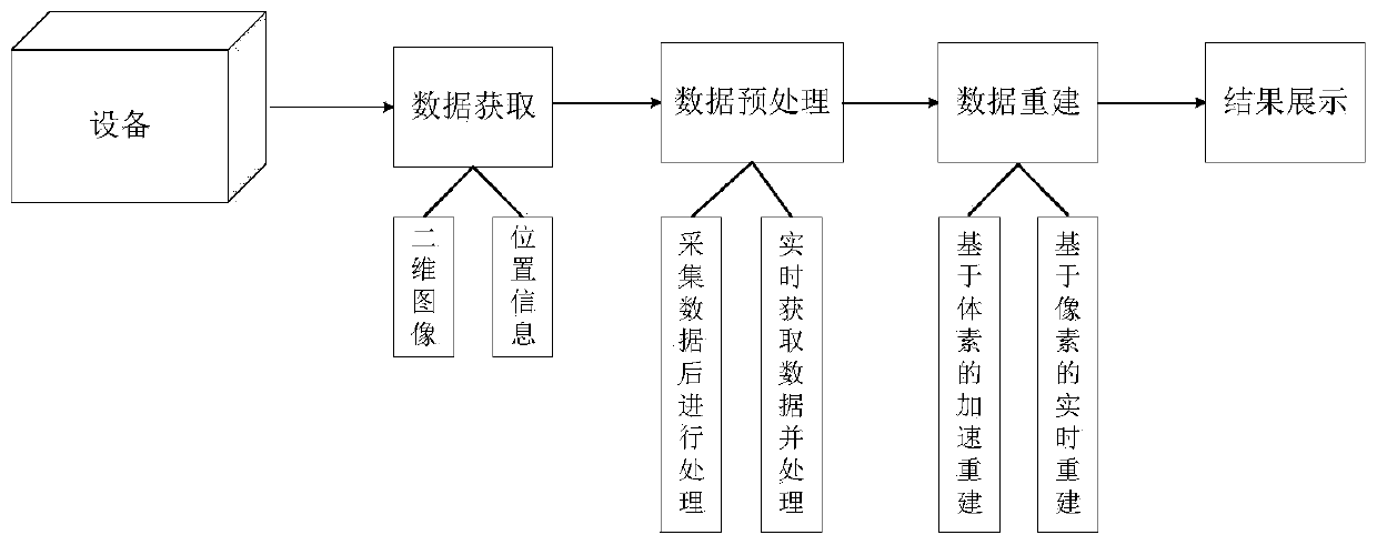
[0057] Such as figure 1 As shown, the present invention provides a kind of application unconstrained scanning and three-dimensional real-time bone imaging method based on voxel, comprising the following steps:

[0058] Step 1: Data Acquisition

[0059] The researcher holds a probe with a position sensor to scan the spine vertically on the back of the human body to obtain a series of two-dimensional cross-sectional ultrasonic ...


PUM


Abstract

The invention provides an unconstrained scanning and voxel-based three-dimensional real-time bone imaging method. According to the method, a voxel-based reconstruction algorithm in a traditional three-dimensional reconstruction technology is applied to skeletal system imaging, a traditional projection method used in the algorithm is optimized and accelerated, and an FDP method is proposed to achieve a real-time reconstruction effect. Because the method provided by the invention is a voxel-based reconstruction algorithm, large-area and large-range imaging (covering the whole back area of a general adult) can be carried out. The method is different from a traditional stacked scanning reconstruction method for three-dimensional bone imaging to the greatest extent. Finally, the technology provided by the invention is also suitable for general non-real-time reconstruction, and is used for accelerating the whole reconstruction process.

Description

technical field [0001] The invention relates to a three-dimensional real-time bone imaging method, which belongs to the technical fields of medical imaging and ultrasonic bone imaging. Background technique [0002] Traditional two-dimensional ultrasound imaging is an indispensable technology in modern medical imaging due to its flexibility, non-invasiveness, no radiation and low cost. However, due to the lack of 2D image information and the difficulty of identification, 3D ultrasound imaging is developing into a new imaging technology. [0003] Three-dimensional ultrasound imaging technology is to embed a sensor in the traditional ultrasound probe to track the position of the probe when it is moved, and then combine the two-dimensional image of the probe to reconstruct a three-dimensional image of the scanned area. At present, in the process of large-scale three-dimensional ultrasound imaging, researchers often use a hand-held probe to scan the region of interest. In the t...

Claims


Application Information

Patent Timeline
Patent Type & Authority Applications(China)
IPC IPC(8): G06T17/00G06T11/00
CPCG06T17/00G06T11/006G06T2207/10136G06T2207/30012
Inventor 郑锐陈宏博
Owner SHANGHAI TECH UNIV
Why Eureka
Try Eureka