CT image-based rib segmentation method, device medium and electronic equipment

A CT image and image technology, applied in the field of image processing, to achieve the effect of reducing workload, improving efficiency, and improving accuracy

Inactive Publication Date: 2020-04-10
INFERVISION MEDICAL TECH CO LTD
View PDF8 Cites 11 Cited by
  • Summary
  • Abstract
  • Description
  • Claims
  • Application Information

AI Technical Summary

Problems solved by technology

[0004] In order to solve the above technical problems, this application proposes a CT image-based rib segmentation method, detection device, computer-readable storage medium and electronic equipment, by obtaining the position information of each pixel in the CT image, and combining the position information with The CT image is input into the neural network model, and the neural network model is used to automatically generate the image of each rib and the corresponding number, thereby greatly reducing the workload of medical staff in the radiology department, improving the efficiency of rib segmentation, and avoiding the error of manual segmentation. situation, improving the accuracy of rib segmentation

Method used


Image

  • CT image-based rib segmentation method, device medium and electronic equipment
  • CT image-based rib segmentation method, device medium and electronic equipment
  • CT image-based rib segmentation method, device medium and electronic equipment

Examples


Embodiment Construction

[0029] Hereinafter, exemplary embodiments according to the present application will be described in detail with reference to the accompanying drawings. Apparently, the described embodiments are only some of the embodiments of the present application, rather than all the embodiments of the present application. It should be understood that the present application is not limited by the exemplary embodiments described here.

[0030] Application overview

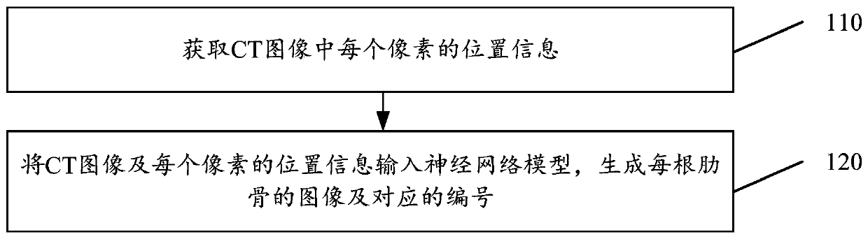
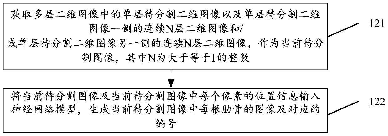
[0031] CT tomographic images are composed of multiple two-dimensional images stacked, which have three-dimensional characteristics. CT tomographic images are an important means and basis for judging whether there are fractures or other traumatic symptoms, such as rib fractures or rib injuries. However, before judging whether there is a fracture, it is necessary to segment each rib from the CT tomographic image and number each rib or each pair of ribs, so as to ensure whether the rib is fractured and the specific number of the f...


PUM


Abstract

The invention discloses a CT image-based rib segmentation method, a CT image-based rib segmentation device, a detection device, a computer readable storage medium and electronic equipment. According to the method, the position information of each pixel in a CT image is acquired; the position information and the CT image are inputted into a neural network model; the image of each rib and the corresponding serial number of the image are automatically generated by utilizing the neural network model; and therefore, the workload of medical personnel in the radiology department is greatly reduced, the efficiency of rib segmentation is improved, the situation of wrong manual segmentation number can be avoided, and the accuracy of the rib segmentation is improved.

Description

technical field [0001] The present application relates to the field of image processing, in particular to a CT image-based rib segmentation method, detection device, computer-readable storage medium and electronic equipment. Background technique [0002] Computed Tomography (CT for short) is a three-dimensional radiological medical image reconstructed after digital geometry processing. This technology mainly irradiates the human body through the rotation of X-rays on a single axis. Since different tissues have different absorption capabilities (or rejection rates) of X-rays, the three-dimensional technology of the computer can be used to reconstruct the tomographic image. By bit processing, the tomographic image of the corresponding tissue can be obtained, and the tomographic image can be stacked layer by layer to form a three-dimensional image. [0003] Human CT tomography is an important means for judging fractures in hospitals, and when rib fractures and lesions are foun...

Claims


Application Information

Patent Timeline
Patent Type & Authority Applications(China)
IPC IPC(8): G06T7/11G06N3/08
CPCG06N3/08G06T7/11G06T2207/10081G06T2207/20081G06T2207/20084G06T2207/30008
Inventor 李玉才陈宽王少康
Owner INFERVISION MEDICAL TECH CO LTD
Why Eureka
Try Eureka