Blood vessel image segmentation method and device, electronic equipment and storage medium

A blood vessel image and blood vessel technology, applied in electronic equipment and storage medium, blood vessel image segmentation method, and device field, can solve problems such as manual selection of growth points

Active Publication Date: 2020-01-17
SHANGHAI MICROPORT PROPHECY MEDICAL TECH CO LTD
View PDF8 Cites 22 Cited by
  • Summary
  • Abstract
  • Description
  • Claims
  • Application Information

AI Technical Summary

Problems solved by technology

[0005] The object of the present invention is to provide a blood vessel image segmentation method, device, electronic equipment and storage medium to solve the problem in the prior art that the growth point must be manually selected when performing blood vessel image segmentation

Method used


Image

  • Blood vessel image segmentation method and device, electronic equipment and storage medium
  • Blood vessel image segmentation method and device, electronic equipment and storage medium
  • Blood vessel image segmentation method and device, electronic equipment and storage medium

Examples


Embodiment Construction

[0095] The following is attached Figures 1 to 4 and Specific Embodiments A blood vessel image segmentation method, device, electronic equipment and storage medium proposed in the present invention will be further described in detail. Advantages and features of the present invention will be apparent from the following description and claims. It should be noted that the drawings are in a very simplified form and all use imprecise scales, which are only used to facilitate and clearly assist the purpose of illustrating the embodiments of the present invention. In order to make the objects, features and advantages of the present invention more comprehensible, please refer to the accompanying drawings. It should be noted that the structures, proportions, sizes, etc. shown in the drawings attached to this specification are only used to match the content disclosed in the specification, for those who are familiar with this technology to understand and read, and are not used to limit ...


PUM


Abstract

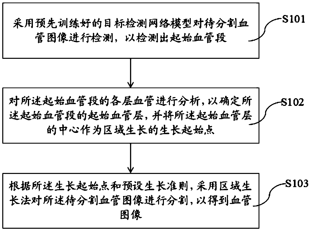
The invention provides a blood vessel image segmentation method and a device, electronic equipment and a storage medium, and the method comprises the steps: employing a pre-trained target detection network model to detect a to-be-segmented blood vessel image, so as to detect an initial blood vessel segment, and the target detection network model is preferably a Faster R-CNN model; analyzing each layer of blood vessel of the initial blood vessel section to determine an initial blood vessel layer of the initial blood vessel section, and taking the center of the initial blood vessel layer as a growth initial point of regional growth; and according to the growth starting point and a preset growth criterion, segmenting the to-be-segmented blood vessel image by adopting a region growth method toobtain a blood vessel image. When the blood vessel image is segmented, the growing points can be automatically calculated, the problem that the growing points need to be manually selected in the prior art is solved, and therefore the segmentation efficiency of the blood vessel image can be effectively improved, and doctors can be better assisted.

Description

technical field [0001] The present invention relates to the technical field of image processing, in particular to a blood vessel image segmentation method, device, electronic equipment and storage medium. Background technique [0002] Aortic aneurysm and aortic dissection are called "time bombs" of the human body. They pose a great threat to human life and health. At present, the commonly used method for diagnosing these two diseases is to perform CT angiography (CTA) on patients to analyze parameters such as vessel shape and diameter. At the same time, it is necessary to perform three-dimensional modeling of aortic vessels on CTA images, and the most critical step in three-dimensional modeling It is blood vessel image segmentation. [0003] Although there are some methods for vascular image segmentation, most of them are done by manual interactive software, such as Mimics software, which requires a lot of manual setting and point selection, and there are some semi-automati...

Claims


Application Information

Patent Timeline
Patent Type & Authority Applications(China)
IPC IPC(8): G06T7/187G06T7/12G06T5/00G06N3/04
CPCG06T7/187G06T7/12G06T5/002G06T2207/10081G06T2207/20036G06T2207/30101G06N3/045
Inventor 张武龙吕文尔黄弯弯
Owner SHANGHAI MICROPORT PROPHECY MEDICAL TECH CO LTD
Why Eureka
Try Eureka