
Medical image segmentation method, device and equipment and storage medium

A medical image and segmentation algorithm technology, applied in the field of medical image processing, can solve the problems of high labor cost and time cost, poor generalization ability of pathological images, over-segmentation and under-segmentation, etc., achieve low labor cost and time cost, improve Segmentation performance, the effect of good segmentation performance

Active Publication Date: 2020-05-12
INFERVISION MEDICAL TECH CO LTD
View PDF10 Cites 9 Cited by
  • Summary
  • Abstract
  • Description
  • Claims
  • Application Information

AI Technical Summary

Problems solved by technology

However, the traditional image segmentation algorithm has the disadvantages of low segmentation accuracy, easy to cause over-segmentation and under-segmentation, and poor generalization ability to pathological images of different tissues; while the deep learning algorithm requires a large amount of artificially labeled perfect data for model training , especially for the cell nucleus segmentation task, which requires fine annotation at the pixel level, which makes the labor cost and time cost of obtaining the perfect data manually annotated higher

Method used


Image

  • Medical image segmentation method, device and equipment and storage medium
  • Medical image segmentation method, device and equipment and storage medium
  • Medical image segmentation method, device and equipment and storage medium

Examples


Embodiment 1

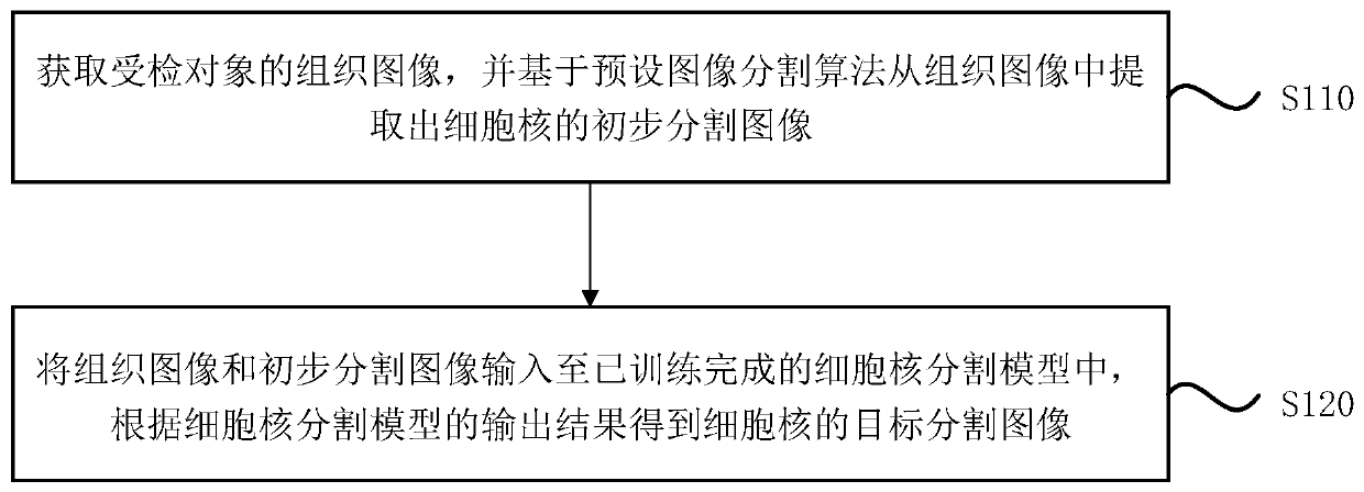
[0042] figure 1 It is a flowchart of a medical image segmentation method provided in Embodiment 1 of the present invention. This embodiment is applicable to the situation of segmenting the cell nucleus image from the tissue image, and is especially applicable to the situation of combining the traditional image segmentation algorithm and the deep learning algorithm to segment the cell nucleus image from the tissue image. The method can be executed by the medical image segmentation device provided by the embodiment of the present invention, the device can be realized by software and / or hardware, and the device can be integrated on various devices.

[0043] see figure 1 , the method of the embodiment of the present invention specifically includes the following steps:

[0044] S110. Acquire a tissue image of the subject to be examined, and extract a preliminary segmented image of cell nuclei from the tissue image based on a preset image segmentation algorithm.

[0045] Wherein,...

Embodiment 2

[0062] Figure 4 It is a structural block diagram of a medical image segmentation device provided in Embodiment 2 of the present invention, and the device is used to implement the medical image segmentation method provided in any of the above embodiments. The device and the medical image segmentation method of the above-mentioned embodiments belong to the same inventive concept. For details not described in detail in the embodiments of the medical image segmentation device, reference can be made to the above-mentioned embodiments of the medical image segmentation method. see Figure 4 , the device may specifically include: a preliminary segmented image extraction module 210 and a target segmented image obtaining module 220 .

[0063] Wherein, the preliminary segmented image extraction module 210 is used to acquire the tissue image of the object under inspection, and extract the preliminary segmented image of cell nuclei from the tissue image based on a preset image segmentati...

Embodiment 3

[0084] Figure 5 A schematic structural diagram of a device provided in Embodiment 3 of the present invention, such as Figure 5 As shown, the device includes a memory 310 , a processor 320 , an input device 330 and an output device 340 . The number of processors 320 in the device may be one or more, Figure 5 Take a processor 320 as an example; the memory 310, processor 320, input device 330 and output device 340 in the device can be connected by bus or other methods, Figure 5 Take the connection via bus 350 as an example.

[0085] The memory 310, as a computer-readable storage medium, can be used to store software programs, computer-executable programs and modules, such as program instructions / modules corresponding to the medical image segmentation method in the embodiment of the present invention (for example, the medical image segmentation device in the Preliminary segmented image extraction module 210 and target segmented image acquisition module 220). The processor ...


PUM


Abstract

The embodiment of the invention discloses a medical image segmentation method and device, equipment and a storage medium. The method comprises the following steps: acquiring a tissue image of a detected object, and extracting a preliminary segmentation image of a cell nucleus from the tissue image based on a preset image segmentation algorithm; and inputting the tissue image and the preliminary segmentation image into a trained cell nucleus segmentation model, and obtaining a target segmentation image of the cell nucleus according to an output result of the cell nucleus segmentation model. According to the embodiment of the invention, a traditional image segmentation algorithm and a deep learning algorithm are combined; the good segmentation performance of the deep learning algorithm can be brought into full play, the requirement of a deep learning algorithm for manually annotated perfect data can be greatly relieved, so that good segmentation performance can still be achieved under the condition that only a small amount of manually annotated perfect data exists, and the effect of accurately segmenting the cell nucleus image from the medical image with low labor cost and time costis achieved.

Description

technical field [0001] Embodiments of the present invention relate to the technical field of medical image processing, and in particular, to a medical image segmentation method, device, equipment, and storage medium. Background technique [0002] As an important part of pathology, histopathological images are an important reference factor for medical staff to diagnose and prognose various diseases. In the analysis of histopathological images, the different shapes of cell nuclei are an important basis for the occurrence of many diseases. Therefore, how to accurately extract the segmented images of cell nuclei from histopathological images is an important topic for computer-aided diagnosis and automatic medical image analysis. . [0003] Medical image segmentation is the process of segmenting medical images into several regions with similar properties, and it is a pixel-by-pixel classification task. For the cell nucleus segmentation task in medical image segmentation, existi...

Claims


Application Information

Patent Timeline
IPC IPC(8): G06T7/194G06T7/187G06T7/155G06T5/00G06N3/08G06N3/04
CPCG06T7/187G06T7/194G06N3/08G06T7/155G06T2207/10056G06T2207/20081G06T2207/20084G06T2207/20016G06T2207/30024G06N3/045G06T5/70Y02A90/10
Inventor 康清波张荣国李新阳陈宽王少康
Owner INFERVISION MEDICAL TECH CO LTD
Learn More