Retinal vessel segmentation method based on symmetric bidirectional cascade network deep learning

A retinal blood vessel and cascade network technology is applied in the field of retinal blood vessel segmentation based on deep learning of symmetric bidirectional cascade network, which can solve the problems of high false positive rate and poor fundus image segmentation effect, achieve a good training process and save calculation. time, the effect of avoiding computational redundancy

Pending Publication Date: 2020-05-15
SHANDONG UNIV OF SCI & TECH
View PDF5 Cites 8 Cited by
  • Summary
  • Abstract
  • Description
  • Claims
  • Application Information

AI Technical Summary

Problems solved by technology

This method has a good segmentation effect on healthy fundus images, but it is not effective

Method used


Image

  • Retinal vessel segmentation method based on symmetric bidirectional cascade network deep learning
  • Retinal vessel segmentation method based on symmetric bidirectional cascade network deep learning
  • Retinal vessel segmentation method based on symmetric bidirectional cascade network deep learning

Examples


Embodiment Construction

[0042] The specific embodiment of the present invention will be further described below in conjunction with accompanying drawing and specific embodiment:

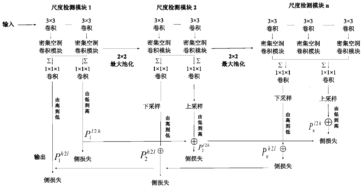
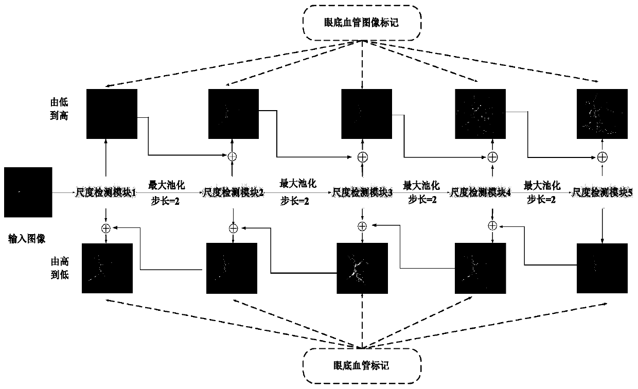
[0043] combine figure 1 and figure 2 , a retinal vessel segmentation method based on symmetric bidirectional cascaded network deep learning, comprising the following steps:

[0044] Step 1: Preprocess the fundus retinal image, and perform data enhancement on the input color fundus original image by cutting, changing contrast, rotating, zooming and translating to realize data set expansion. The specific steps include:

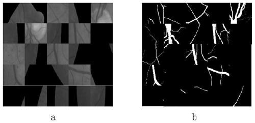
[0045] Step 1.1: Divide the fundus retinal image and its corresponding Groundtruth (standard blood vessel segmentation map) into 50×50 slices. The original image slices in the STARE dataset and the corresponding Groundtruth (standard blood vessel segmentation map) slices are as follows image 3 shown in a and 3b. For the STARE dataset, only one retinal image-generated slice is tested at a time, while th...


PUM


Abstract

The invention discloses a retinal vessel segmentation method based on symmetric bidirectional cascade network deep learning, and belongs to the field of medical image processing. According to the method, data enhancement is carried out through a series of modes such as contrast change, rotation, zooming and translation, data set amplification is realized, and then a preprocessed image is input into a bidirectional cascade network for training and learning to obtain a predicted retinal vessel segmentation result. The network is composed of five scale detection modules, retinal vessel characteristics of different diameter scales are extracted by changing the expansion rate, two vessel contour prediction images are generated in the two directions from the lower layer to the upper layer and from the upper layer to the lower layer of the network respectively, and the two vessel contour prediction images are structurally distributed in an up-and-down symmetrical mode; the outputs of the twopaths of the dense hole convolution module are combined; and finally, the blood vessels and the background pixels are classified by adopting a quasi-balanced cross entropy loss function so as to realize accurate segmentation of the retinal blood vessels.

Description

technical field [0001] The invention belongs to the field of medical image processing, and in particular relates to a retinal blood vessel segmentation method based on deep learning of a symmetrical bidirectional cascaded network. Background technique [0002] Retinal blood vessels are the only clear blood vessels in the human body that can be observed by non-invasive means. Current medical research shows that retinal vascular abnormalities not only manifest as eye diseases such as glaucoma and cataract, but are also directly related to the severity of cardiovascular diseases such as hypertension, coronary heart disease, diabetes, and atherosclerosis. The morphological structure of retinal blood vessels in the fundus can reflect the condition of the eye and the vascular system around the body. Through the analysis of retinal images, it can effectively predict, diagnose and prevent cardiovascular diseases. Therefore, the research on blood vessel segmentation technology based...

Claims


Application Information

Patent Timeline
IPC IPC(8): G06T7/12G06T7/194G06T7/00G06N3/04G06N3/08
CPCG06T7/12G06T7/194G06T7/0012G06N3/08G06T2207/30041G06N3/045
Inventor 彭延军郭燕飞王元红
Owner SHANDONG UNIV OF SCI & TECH
Why Eureka
Try Eureka