Medical imaging method and medical imaging method equipment
A technology of medical imaging and equipment, applied in the field of medical imaging, can solve problems such as unclear explanation or discussion, and achieve the effects of easy disassembly and analysis, clear imaging, and improved work efficiency
- Summary
- Abstract
- Description
- Claims
- Application Information
AI Technical Summary
Problems solved by technology
Method used
Image

Examples
Embodiment 1
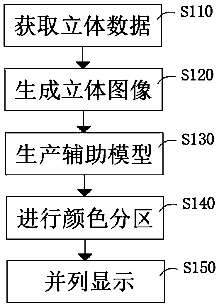
[0044] Refer to attached Figure 1-5 Shown, a kind of medical imaging method of the present embodiment, imaging method comprises the following steps:
[0045] S110, acquiring stereoscopic data of the part to be detected;
[0046] S120, generating a stereoscopic image according to the data;
[0047] S130, generating an auxiliary model according to the stereo image;
[0048] S140, performing color partitioning based on the auxiliary model;
[0049] S150, display the stereoscopic image and the auxiliary model side by side.
[0050] The stereoscopic image in this embodiment is the internal structure diagram of the part detected at that time, and the stereoscopic image is a black and white image with different brightness and darkness.
[0051] The auxiliary model in this embodiment is generated based on a stereoscopic image, and the auxiliary model divides the different light and dark contents in the stereoscopic image into editable regions, and the name, color and status of th...
Embodiment 2
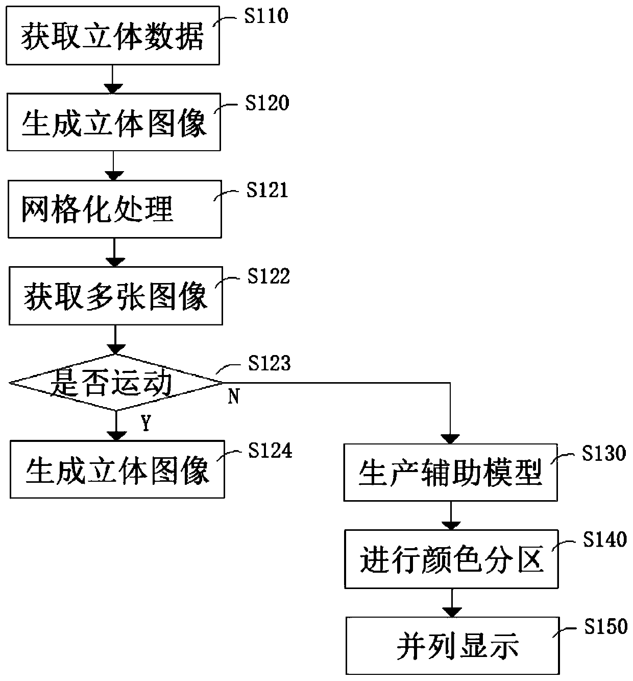
[0053] Refer to attached Figure 1-5 Shown, a kind of medical imaging method of the present embodiment, imaging method comprises the following steps:
[0054] S110, acquiring stereoscopic data of the part to be detected;
[0055] S120, generating a stereoscopic image according to the data;
[0056] S130, generating an auxiliary model according to the stereo image;
[0057] S140, performing color partitioning based on the auxiliary model;
[0058] S150, display the stereoscopic image and the auxiliary model side by side.
[0059] The stereoscopic image in this embodiment is the internal structure diagram of the part detected at that time, and the stereoscopic image is a black and white image with different brightness and darkness.
[0060] The auxiliary model in this embodiment is generated based on a stereoscopic image, and the auxiliary model divides the different light and dark contents in the stereoscopic image into editable regions, and the name, color and status of th...
Embodiment 3
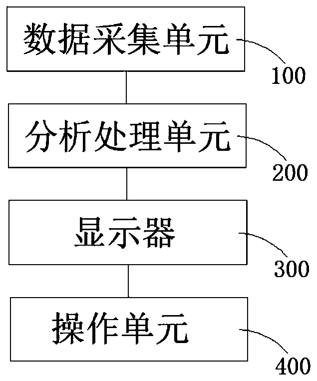
[0069] Refer to attached Figure 1-5 As shown, a kind of medical imaging equipment of the present embodiment comprises:
[0070] A data acquisition unit 100, for acquiring stereoscopic image information of an object;
[0071] The processing and analysis unit 200, processing and analyzing the stereoscopic image to generate an auxiliary model;
[0072] Display 300, displaying stereoscopic images and auxiliary models;
[0073] The operation unit 400 is used to edit the auxiliary model.
[0074] The stereoscopic image acquired by the medical imaging device of this embodiment is an ultrasonic detection image or a nuclear magnetic resonance image.
[0075] The medical imaging device in this embodiment acquires several time-continuous plane coordinate images with a time interval of less than 0.1S.
PUM
 Abstract
Description
Claims
Application Information
 - Generate Ideas
- Intellectual Property
- Life Sciences
- Materials
- Tech Scout
- Unparalleled Data Quality
- Higher Quality Content
- 60% Fewer Hallucinations
Browse by: Latest US Patents, China's latest patents, Technical Efficacy Thesaurus, Application Domain, Technology Topic, Popular Technical Reports.
© 2025 PatSnap. All rights reserved.Legal|Privacy policy|Modern Slavery Act Transparency Statement|Sitemap|About US| Contact US: help@patsnap.com