Brain lesion image spatial distribution characteristic classification and identification method based on magnetic resonance imaging

A magnetic resonance imaging and distribution feature technology, applied in image analysis, image enhancement, image data processing, etc., can solve the problems of insufficient objectivity, limited features, and low efficiency

Pending Publication Date: 2020-07-14
AFFILIATED HUSN HOSPITAL OF FUDAN UNIV
View PDF2 Cites 7 Cited by
  • Summary
  • Abstract
  • Description
  • Claims
  • Application Information

AI Technical Summary

Problems solved by technology

This method of qualitative identification and counting by doctors, on the one hand, the extracted features are extremely limited, and it is not objective enough, and there are differences in the discrimination results of different doctors or researchers; on the other hand, it has not been automated and the efficiency is low

Method used


Image

  • Brain lesion image spatial distribution characteristic classification and identification method based on magnetic resonance imaging
  • Brain lesion image spatial distribution characteristic classification and identification method based on magnetic resonance imaging
  • Brain lesion image spatial distribution characteristic classification and identification method based on magnetic resonance imaging

Examples


Embodiment 1

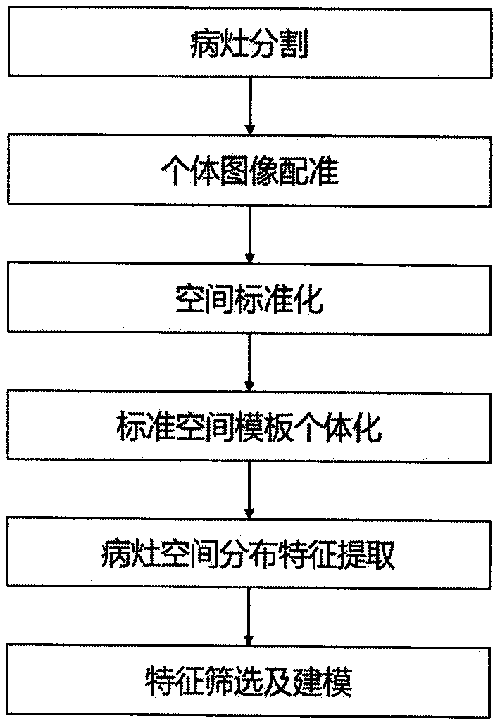
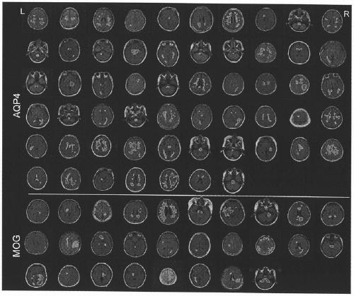
[0036] Example 1 Classification and identification of spatial distribution characteristics of brain lesion images of MOG antibody-positive and AQP4 antibody-positive NMOSD patients

[0037] Classification identification:

[0038] 1) Select the clinical MRI images of MOG antibody-positive and AQP4 antibody-positive NMOSD groups, 28 cases and 57 cases respectively, and each case includes FLAIR images as "lesion display images" and T1-weighted images as "brain structure images" ;

[0039] 2) For the FLAIR image, use MRIcron to segment the whole brain lesion and save it as a binary image as a "lesion image";

[0040] 3) Use SPM to rigidly register the T1-weighted image of the individual with the FLAIR image;

[0041] 4) Use SPM to register the individual T1-weighted image to the MNI standard space; apply the transformation parameters to the FLAIR image, and select a threshold of 0.5 to obtain a binarized "standard space lesion image";

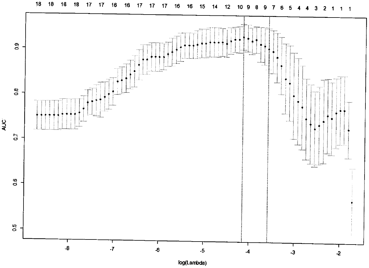
[0042] 5) Use the individual space "lesio...


PUM


Abstract

The invention belongs to the technical field of image processing and application, and particularly relates to a brain lesion image spatial distribution feature classification and identification methodbased on magnetic resonance imaging. The method mainly comprises the steps of focus segmentation, individual image registration, space standardization, standard space template individualization, focus space distribution feature extraction, feature screening, modeling and the like. The core is in that an analysis method of a brain lesion image spatial distribution feature set is constructed by analyzing various features of lesions in an individual space and a standard space, and feature screening and modeling are carried out by using machine learning on the basis of the analysis method. The method can be used for classifying and identifying brain lesion images of different brain diseases or brain states caused by different antibodies, different genes and the like by using brain magnetic resonance images, and effective guidance is provided for clinic and scientific research.

Description

technical field [0001] The invention belongs to the field of image processing and application, and in particular relates to a method for classifying and identifying spatial distribution characteristics of brain lesion images based on magnetic resonance imaging. Background technique [0002] In the prior art, in the classification and identification methods of different brain diseases or brain state images, brain magnetic resonance imaging technology plays an important role due to its non-invasiveness, timeliness, and excellent display effect on brain lesions. In clinical practice, doctors often use long-term clinical experience to summarize the different characteristics of lesions of different diseases on images, and perform visual classification, identification and reporting. However, this experience-based classification and identification has shortcomings such as low efficiency, difficulty in finding new features of lesions, and difficulty in automatically combining multip...

Claims


Application Information

Patent Timeline
Patent Type & Authority Applications(China)
IPC IPC(8): G06T7/00G06T7/136G06K9/62
CPCG06T7/0012G06T7/136G06T2207/10088G06T2207/30016G06T2207/20084G06T2207/20104G06F18/2411
Inventor 杨丽琴夏威李郁欣耿道颖李海庆
Owner AFFILIATED HUSN HOSPITAL OF FUDAN UNIV
Why Eureka
Try Eureka