
Chest X-ray disease detection device and method based on dual-channel separation network

A technology for disease detection and separation of networks, applied in biological neural network models, instruments for radiological diagnosis, image analysis, etc. It can solve problems such as lack of annotations/labels for training datasets, poor supervision of noisy labels, and difficulty in detecting diseased areas. , to improve the quality of medical services, promote information sharing and integration, and facilitate secondary diagnosis.

Active Publication Date: 2020-07-17
SHENZHEN GRADUATE SCHOOL TSINGHUA UNIV
View PDF4 Cites 9 Cited by
  • Summary
  • Abstract
  • Description
  • Claims
  • Application Information

AI Technical Summary

Problems solved by technology

[0005] 1) Lack of training data sets and related annotations / labels of images, professional knowledge limitations lead to high difficulty in labeling
In addition, even if there is data labeled by experts, the existing label noise will also interfere
[0006] 2) The visual patterns extracted from different types of chest disease samples are usually highly diverse in appearance, size, and location, and traditional image processing methods cannot learn meaningful representations due to poor supervision of noisy labels
[0007] 3) The image category is unbalanced, and there are usually many more normal images than abnormal images in the database, resulting in the clinical features being easily overwhelmed
Local pathological image regions can show different sizes or extents, but are usually very small compared to the full image scale, lesion regions are difficult to detect, and training classifiers on X-ray images is more difficult than general-purpose images

Method used


Image

  • Chest X-ray disease detection device and method based on dual-channel separation network
  • Chest X-ray disease detection device and method based on dual-channel separation network
  • Chest X-ray disease detection device and method based on dual-channel separation network

Examples


Embodiment Construction

[0033] Embodiments of the present invention will be described in detail below. It should be emphasized that the following description is only exemplary and not intended to limit the scope of the invention and its application.

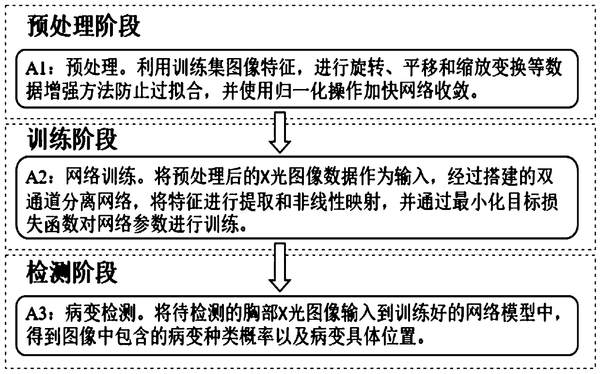
[0034] figure 1 It is a simplified processing flow diagram of a device for detecting diseases in chest X-ray images based on a dual-channel separation network according to an embodiment of the present invention. An embodiment of the present invention provides a chest X-ray disease detection device based on a two-channel separation network, including a processor configured to execute figure 1 Steps A1-A3 shown:

[0035] Step 1: Preprocess the chest X-ray image training data set and divide it into two parts: data enhancement and normalization. This process can expand the training data set, reduce overfitting, accelerate network convergence, and improve network generalization performance .

[0036] In this step, X-ray images containing multiple frontal...


PUM


Abstract

The invention discloses a chest X-ray disease detection device and method based on a dual-channel separation network, and the device comprises a processor which is configured to carry out the following operations: carrying out the preprocessing of a training data set of a chest X-ray image, and dividing the training data set into a data enhancement part and a normalization regularization part; training a dual-channel separation depth network, extracting and fusing features of each level in different channels, minimizing a loss function in a classification layer, and performing complete training of the network; classifying the input chest X-ray images by using the trained network to obtain lesion types and probabilities contained in the images; and using the trained network to locate the disease in the input chest X-ray image. The method can greatly improve the recognition accuracy of chest lesions in a chest disease recognition task, and can achieve the precise positioning of a diseaseposition in a visualization task.

Description

technical field [0001] The invention relates to the fields of computer vision and medical image processing, in particular to a chest X-ray disease detection device and method based on a dual-channel separation network. Background technique [0002] Medical X-ray images are an imaging method that reflects the internal structure of the human body according to the different absorption levels of X-rays by different tissues, and can display a variety of complex pathological features. Chest X-ray (CXR) imaging is one of the most widely available radiological examinations for screening and clinical diagnosis. However, automatic detection and understanding of CXR images has become a technically challenging task at present due to the complex pathology of different types of lung lesions on the images. Because chest X-ray imaging technology is not only fast, simple, but also economical, people usually choose X-ray when checking chest diseases such as thorax (including ribs, thoracic s...

Claims


Application Information

Patent Timeline
IPC IPC(8): G06T7/00G06K9/62G06N3/04A61B6/00
CPCG06T7/0012A61B6/00A61B6/52G06T2207/10116G06T2207/20081G06T2207/20084G06T2207/30004G06V2201/03G06N3/045G06F18/213G06F18/2415G06F18/253
Inventor 王好谦胡小婉张永兵
Owner SHENZHEN GRADUATE SCHOOL TSINGHUA UNIV
Learn More