
Medical ultrasonic image segmentation method and device

An ultrasonic image and medical technology, applied in the field of medical ultrasonic image segmentation method and device, can solve problems such as insufficient segmentation accuracy, inaccurate segmentation, and unsatisfactory segmentation effect, and achieve the effect of improving anti-interference performance and improving segmentation accuracy

Pending Publication Date: 2020-07-17
深圳市嘉骏实业有限公司
View PDF3 Cites 4 Cited by
  • Summary
  • Abstract
  • Description
  • Claims
  • Application Information

AI Technical Summary

Problems solved by technology

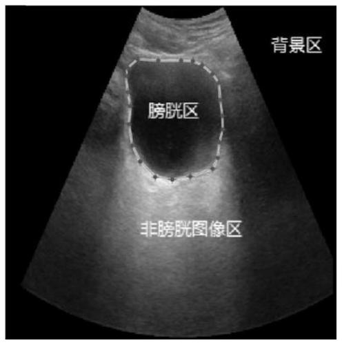
[0004] In the prior art, there have been many bladder image segmentation methods, but the existing bladder ultrasound image segmentation, because there are many artifacts in the bladder ultrasound image, the anterior wall of the bladder often has brighter multiple reflection signals, resulting in a simple The threshold-based segmentation method is difficult to completely segment the bladder area
Region-based segmentation methods are also affected by this artifact and lead to inaccurate segmentation
In addition, in bladder ultrasound images, on the bladder side wall, due to the incident angle and attenuation of the ultrasound signal, the image signal-to-noise ratio is very low, and the algorithm based on edge detection is not ideal for segmentation in such a low signal-to-noise ratio image
Based on the method of deep learning, the accuracy of segmentation is relatively high, but the segmentation accuracy is not high enough, especially the segmented bladder boundary, which often cannot completely fit the edge of the bladder wall

Method used


Image

  • Medical ultrasonic image segmentation method and device
  • Medical ultrasonic image segmentation method and device
  • Medical ultrasonic image segmentation method and device

Examples


Embodiment Construction

[0043] The following will clearly and completely describe the technical solutions in the embodiments of the present invention with reference to the accompanying drawings in the embodiments of the present invention. Obviously, the described embodiments are some of the embodiments of the present invention, but not all of them. Based on the embodiments of the present invention, all other embodiments obtained by persons of ordinary skill in the art without making creative efforts belong to the protection scope of the present invention.

[0044] It should be understood that when used in this specification and the appended claims, the terms "comprising" and "comprises" indicate the presence of described features, integers, steps, operations, elements and / or components, but do not exclude one or Presence or addition of multiple other features, integers, steps, operations, elements, components and / or collections thereof.

[0045] It should also be understood that the terminology used ...


PUM


Abstract

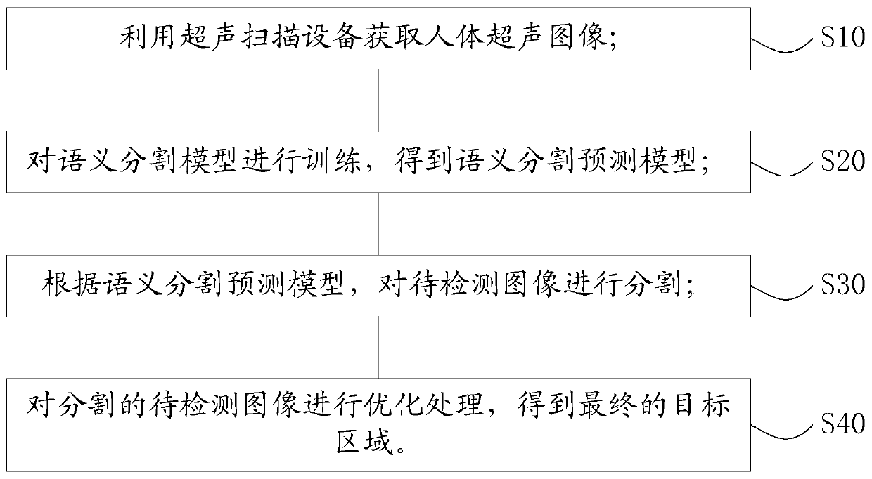
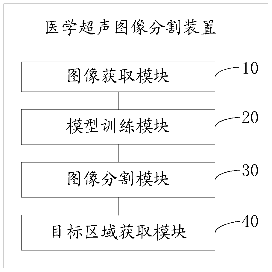
The invention provides a medical ultrasonic image segmentation method and device, and the method comprises the steps: obtaining a human body ultrasonic image through ultrasonic scanning equipment; training the semantic segmentation model to obtain a semantic segmentation prediction model; segmenting the to-be-detected image according to the semantic segmentation prediction model; and performing optimization processing on the segmented to-be-detected image to obtain a final target area. The medical ultrasonic image segmentation method and device have the advantages that a deep learning method and a traditional image processing method are combined, the problem that segmentation is inaccurate when the signal-to-noise ratio of the ultrasonic image is low can be effectively solved, the segmentation precision of the bladder ultrasonic image can be improved, and the anti-interference performance of the algorithm is improved.

Description

technical field [0001] The invention relates to the field of image segmentation, in particular to a medical ultrasound image segmentation method and device. Background technique [0002] Image segmentation plays an important role in imaging diagnosis. Automatic segmentation can help doctors quickly determine the size and location of organs or lesions, and can reduce the heavy workload caused by manual labeling by doctors. [0003] The bladder is a muscular cystic organ that stores urine in the human body. The volume of the bladder reflects the amount of urine that the human body can store, and is also an important parameter for clinical application in urology. In the diagnosis and treatment of the urinary system, the measurement results of the residual urine volume of the human bladder and the filled urine volume of the bladder are important references for doctors to diagnose cases. Non-invasive measurement of bladder volume using ultrasound equipment has been widely used c...

Claims


Application Information

Patent Timeline
Patent Type & Authority Applications(China)
IPC IPC(8): G06T7/00G06T7/11
CPCG06T7/0012G06T7/11G06T2207/10132G06T2207/20081G06T2207/20084G06T2207/30004Y02T10/40
Inventor 张官喜
Owner 深圳市嘉骏实业有限公司
Learn More