A diagnosis system for laryngeal diseases based on deep learning neural network

A technology of disease diagnosis and neural network, which is applied in interdisciplinary fields, can solve the problems of low diagnosis accuracy of laryngoscope image diagnosis, achieve the effect of improving diagnosis efficiency and diagnosis accuracy, good diagnosis, and reducing missed diagnosis and misdiagnosis rate

Active Publication Date: 2020-12-11
HARBIN INST OF TECH
View PDF13 Cites 1 Cited by
  • Summary
  • Abstract
  • Description
  • Claims
  • Application Information

AI Technical Summary

Problems solved by technology

[0006] The purpose of the present invention is to solve the problem of low diagnostic efficiency and diagnostic accuracy of laryngoscope images by traditional methods, and propose a laryngeal disease diagnosis system based on deep learning neural network

Method used


Image

  • A diagnosis system for laryngeal diseases based on deep learning neural network
  • A diagnosis system for laryngeal diseases based on deep learning neural network
  • A diagnosis system for laryngeal diseases based on deep learning neural network

Examples


specific Embodiment approach 1

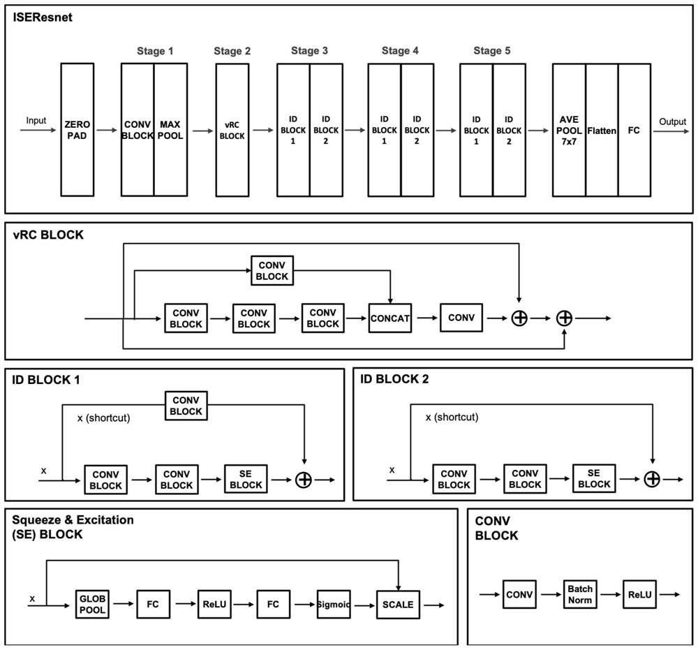
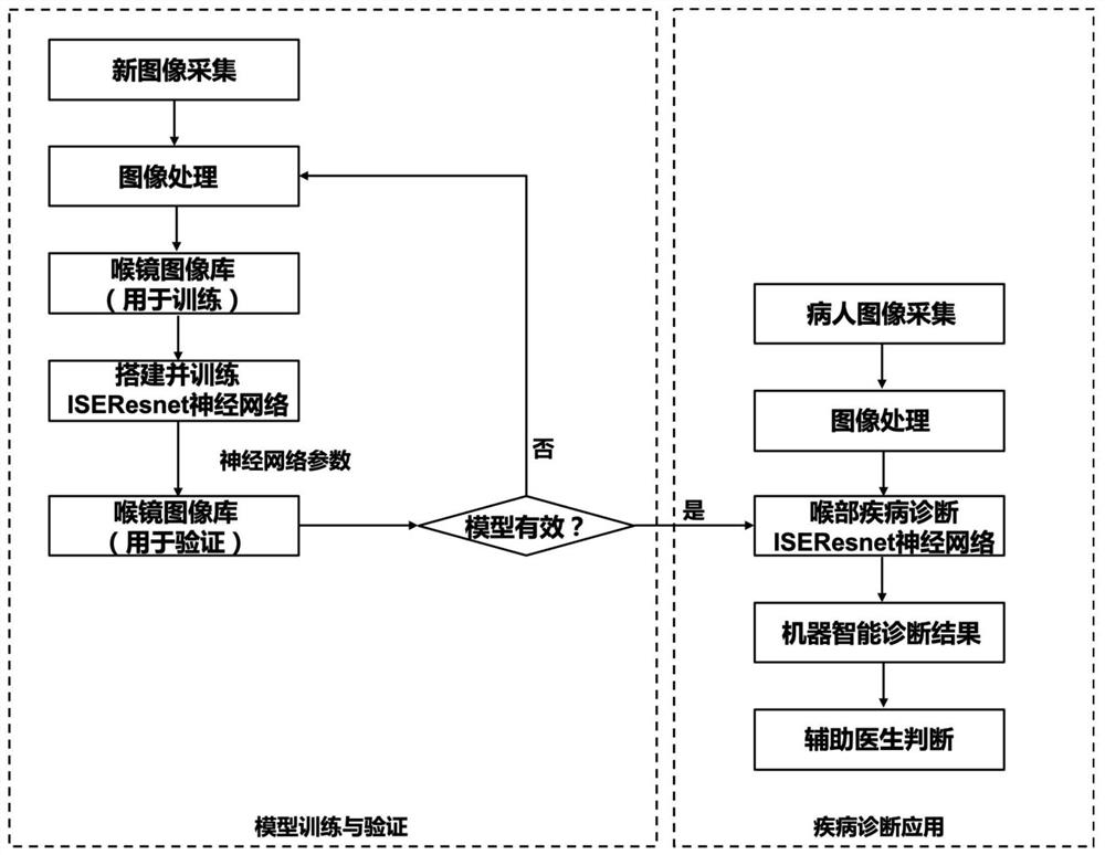
[0020] Specific implementation mode 1. Combination figure 1 This embodiment will be described. A laryngeal disease diagnosis system based on a deep learning neural network described in this embodiment, the laryngeal disease diagnosis system includes an image acquisition main module, an image processing main module, a neural network main module, a training main module and a detection main module ;

[0021] The image collection main module is used to collect laryngoscope images, preprocess the collected laryngoscope images, obtain preprocessed images, and input the preprocessed images into the image processing main module;

[0022] The image processing main module is used to process the input image, and randomly divide the processed image into two groups of training sample set and verification sample set;

[0023] The neural network main module is used to build a network model for laryngeal disease diagnosis;

[0024] The training main module uses the training sample set to t...

specific Embodiment approach 2

[0031] Embodiment 2: The difference between this embodiment and Embodiment 1 is that the image acquisition main module scans the laryngoscope paper image output by the instrument or the laryngoscope paper image attached to the patient's medical record into an electronic The format of the image, after obtaining each complete laryngoscope electronic image, split the 4 sub-images on each image, and adjust the angle of the split image to make the split image correct;

[0032] After the white frame of the corrected image is removed, the image is adjusted to a uniform size; the size-adjusted image is input into the image processing main module.

specific Embodiment approach 3

[0033] Embodiment 3: The difference between this embodiment and Embodiment 1 is that the image processing main module is used to process the input image, and the specific process of processing is as follows:

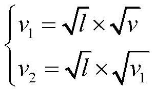
[0034] Perform HSV decomposition on each image input to the image processing main module, wherein H, S and V represent the hue, saturation and brightness of the image respectively;

[0035] Do the following transformation on the points whose luminance value is greater than the luminance threshold l in the V channel (brightness channel):

[0036]

[0037] Among them, v represents the brightness value in the original channel, and l represents the brightness threshold (that is, when the brightness value v in the original channel is greater than the brightness threshold l, it will be transformed), v 1 is an intermediate variable, v 2 Represents the transformed brightness value;

[0038] Then normalize each image input to the image processing main module, so that the val...


PUM


Abstract

A laryngeal disease diagnosis system based on deep learning neural network, which belongs to the interdisciplinary field of artificial intelligence and medical diagnosis. The invention solves the problem of low diagnostic efficiency and diagnostic accuracy of the laryngoscope image by the traditional method. The present invention builds a laryngeal disease diagnosis network model, and the built laryngeal disease diagnosis network model can be used in an intelligent system for laryngeal disease diagnosis, thereby better diagnosing laryngoscope images and helping doctors improve disease diagnosis efficiency and diagnosis Accuracy, reduce missed diagnosis and misdiagnosis rate. The invention can be applied to intelligent detection of laryngoscope images.

Description

technical field [0001] The invention relates to the interdisciplinary field of combining artificial intelligence and medical diagnosis, in particular to a laryngeal disease diagnosis system based on a deep learning neural network. Background technique [0002] Due to the special position and complex physiological structure of the human throat, it is often impossible to directly spy on it. When diagnosing diseases of the larynx, doctors often obtain internal information by inserting a laryngoscope into the larynx to take images, and then carry out diagnosis and treatment. In clinical practice, fiberoptic laryngoscopy is often used for diagnosis and treatment. Fiber laryngoscope is a kind of fiber optic device, which causes less trauma and helps to reduce the pain of patients. It can also enlarge the lesion site through fiber imaging technology, provide a clear field of view, and help doctors make better judgments. [0003] Laryngoscopy can usually be used for initial screen...

Claims


Application Information

Patent Timeline
Patent Type & Authority Patents(China)
IPC IPC(8): G06K9/62G06N3/04G06N3/08G16H50/20
CPCG06N3/08G16H50/20G06N3/045G06F18/241G06F18/214
Inventor 赵雪岩罗浩刘绍宠刘富豪蒋宇辰尹珅
Owner HARBIN INST OF TECH
Why Eureka
Try Eureka