Image segmentation method and related equipment

An image segmentation and image technology, applied in the field of medical imaging, can solve the problems of inaccurate detection results and inaccurate positioning, and achieve the effect of improving segmentation accuracy and robustness

Pending Publication Date: 2020-08-04
SHANGHAI SENSETIME INTELLIGENT TECH CO LTD
View PDF10 Cites 5 Cited by
  • Summary
  • Abstract
  • Description
  • Claims
  • Application Information

AI Technical Summary

Problems solved by technology

[0003] The traditional segmentation method of the infarct core area and the ischemic penumbra area based on the mathematical model has shortcomings such as inaccurate positioning and inaccurate detection results.

Method used


Image

  • Image segmentation method and related equipment
  • Image segmentation method and related equipment
  • Image segmentation method and related equipment

Examples


Embodiment Construction

[0030] The following will clearly and completely describe the technical solutions in the embodiments of the present application with reference to the accompanying drawings in the embodiments of the present application. Obviously, the described embodiments are only part of the embodiments of the present application, not all of them. Based on the embodiments in this application, all other embodiments obtained by persons of ordinary skill in the art without making creative efforts belong to the scope of protection of this application.

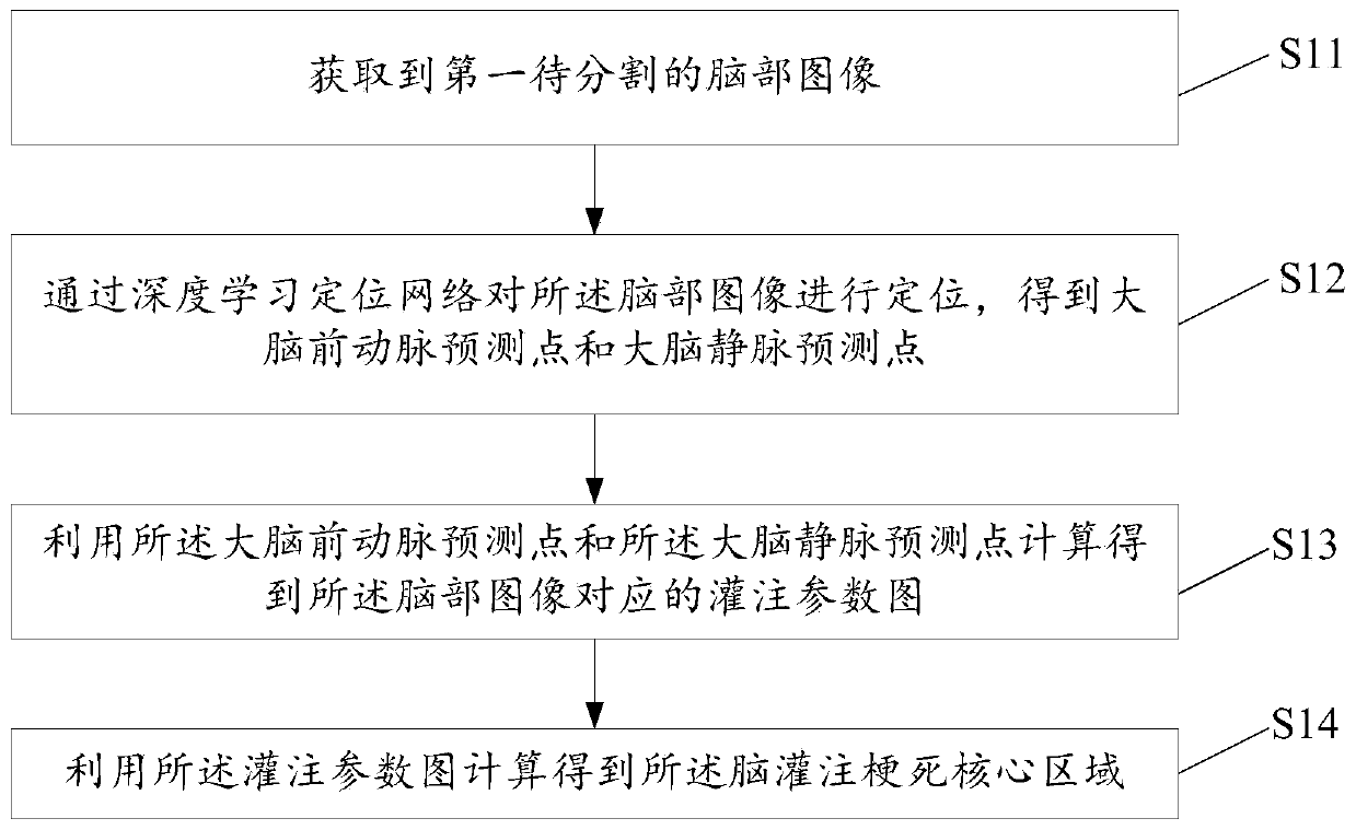
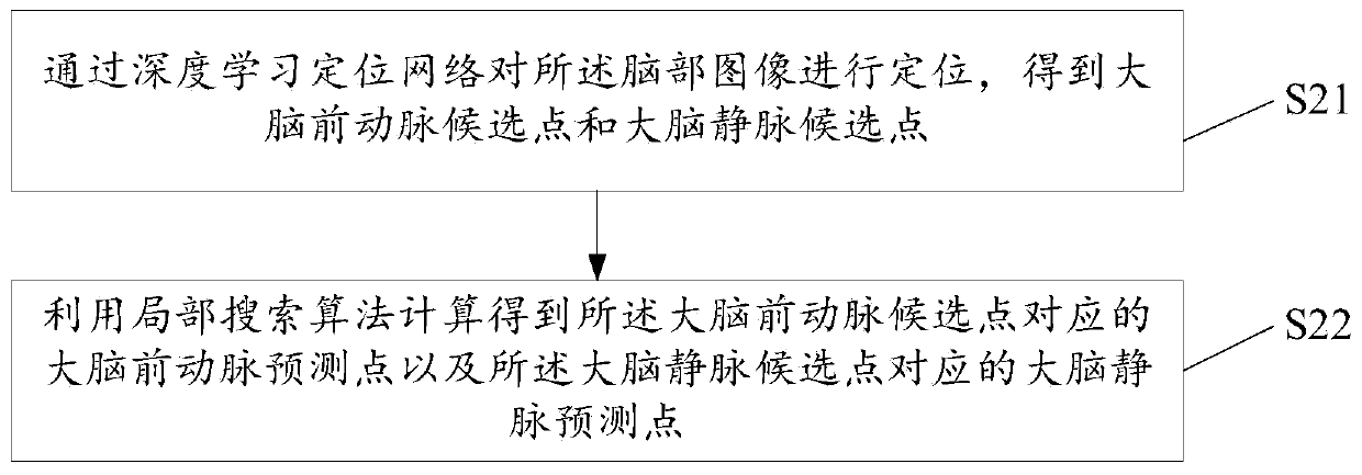
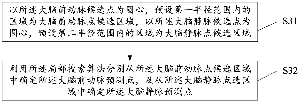
[0031] The image segmentation method provided by the present invention uses a deep learning algorithm to locate the anterior cerebral artery points and cerebral vein points, which can effectively solve the shortcomings of the automatic or manual positioning of the cerebral artery points and vein points in the prior art. Provide protection for subsequent calculations. Moreover, the image segmentation method provided by the present invention does no...


PUM


Abstract

The invention provides an image segmentation method and a related device, and the method comprises the steps: obtaining a first to-be-segmented brain image; positioning the brain image through a deeplearning positioning network to obtain a cerebral anterior artery prediction point and a cerebral vein prediction point; calculating to obtain a perfusion parameter graph corresponding to the brain image by utilizing the cerebral anterior artery prediction point and the cerebral vein prediction point; and calculating to obtain the cerebral perfusion infarction core region by utilizing the perfusion parameter graph, so as to improve the segmentation precision and robustness of the infarction core region.

Description

technical field [0001] The invention relates to the technical field of medical imaging, in particular to an image segmentation method and related equipment. Background technique [0002] Brain CT perfusion imaging is an imaging technique used to analyze intracranial hemodynamics and is widely used in the diagnosis of ischemic stroke. CT perfusion imaging is to observe the dynamic changes of brain tissue density during rapid intravenous bolus injection of iodine contrast agent, and calculate the regional cerebral blood volume, regional cerebral blood flow, average transit time and peak time according to different mathematical models. If the time is longer than 6 seconds, it is a hypoperfusion area. The local cerebral blood flow on the lesion side is less than 30% of the local cerebral blood flow on the upper normal side, which is regarded as the core area of ​​cerebral infarction. In the hypoperfusion area, the rest area except the infarction core area is the ischemic penumbr...

Claims


Application Information

Patent Timeline
Patent Type & Authority Applications(China)
IPC IPC(8): G06T7/11G06T7/00G06N3/04G06N3/08
CPCG06T7/11G06T7/0012G06N3/08G06T2207/10081G06T2207/30016G06N3/045A61B6/5217A61B6/507A61B6/501A61B6/032G06T2207/30104
Inventor 宋涛
Owner SHANGHAI SENSETIME INTELLIGENT TECH CO LTD
Why Eureka
Try Eureka