Medical image three-dimensional reconstruction method based on three-dimensional image segmentation

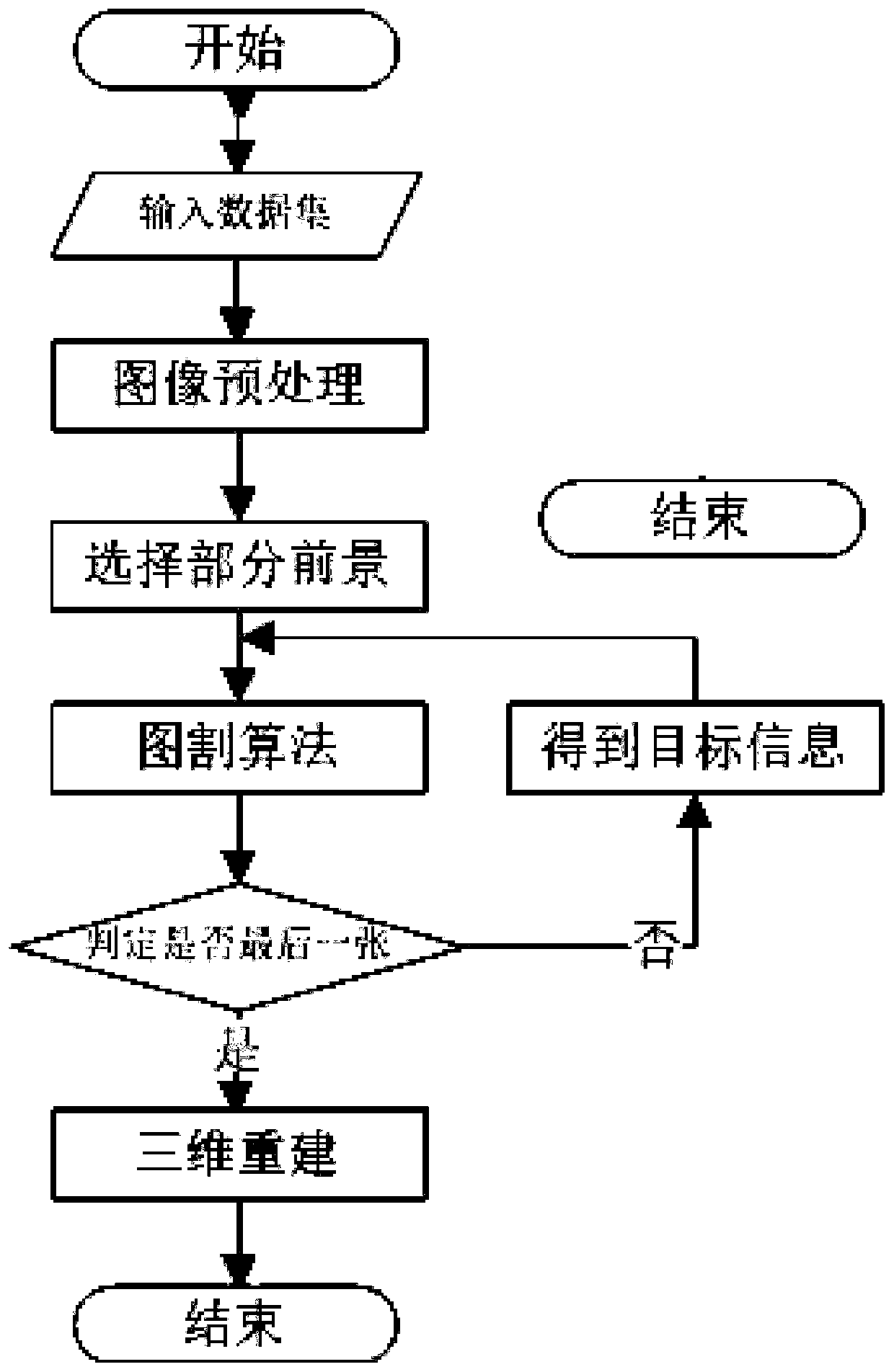
A technology of 3D reconstruction and 3D image, which is applied in the field of 3D reconstruction of medical images based on 3D graph cuts, can solve problems such as low segmentation efficiency, unsatisfactory segmentation results, and poor segmentation effects, so as to ensure accuracy, segmentation efficiency, and good 3D Reconstruction effect, effect of reduced manipulation steps

Pending Publication Date: 2020-08-04
B SOFT CO LTD
View PDF6 Cites 0 Cited by
  • Summary
  • Abstract
  • Description
  • Claims
  • Application Information

AI Technical Summary

Problems solved by technology

[0004] However, the traditional graph cut algorithm only considers the grayscale information of the image. For some complex medical images, the segmentation target and the gray value around the target are very close, resulting in unsatisfactory segmentation results. When applied on the Internet, the algorithm has poor segmentation effect and low segmentation efficiency
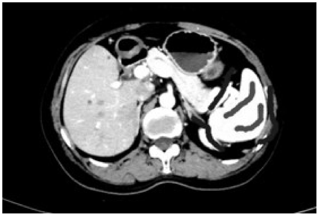
For example, the prior art CN108596887A provides a method for automatically segmenting liver tumors in abdominal CT sequence images. The method is based on the graph cut algorithm to segment liver tumors in abdominal CT sequence images. When the scheme aims to solve the fuzzy boundary of liver tumors in CT images, improve It improves the accuracy and efficiency of computer-aided diagnosis. Although it also realizes the automatic segmentation of CT images, it needs to perform grayscale processing on each medical image, and the efficiency is low in multi-image processing.

Method used


Image

  • Medical image three-dimensional reconstruction method based on three-dimensional image segmentation
  • Medical image three-dimensional reconstruction method based on three-dimensional image segmentation
  • Medical image three-dimensional reconstruction method based on three-dimensional image segmentation

Examples


Embodiment Construction

[0019] The following will clearly and completely describe the technical solutions in the embodiments of the present invention with reference to the accompanying drawings in the embodiments of the present invention. Obviously, the described embodiments are only some, not all, embodiments of the present invention. All other embodiments obtained by persons of ordinary skill in the art based on the embodiments of the present invention belong to the protection scope of the present invention.

[0020] Those skilled in the art should understand that in the disclosure of the present invention, the terms "vertical", "transverse", "upper", "lower", "front", "rear", "left", "right", " The orientation or positional relationship indicated by "vertical", "horizontal", "top", "bottom", "inner", "outer", etc. is based on the orientation or positional relationship shown in the drawings, which are only for the convenience of describing the present invention and The above terms should not be con...


PUM


Abstract

The invention provides a medical image three-dimensional reconstruction method based on three-dimensional image segmentation, and the method improves an image segmentation algorithm according to the structural similarity of continuous medical images, achieves the automatic segmentation of medical images, enables a segmentation result of the images to be used for three-dimensional reconstruction, and achieves a better reconstruction effect.

Description

technical field [0001] The invention relates to the technical field of medical image processing, in particular to a three-dimensional reconstruction method of medical images based on three-dimensional graph cuts. Background technique [0002] Medical imaging refers to the technology and processing process of obtaining internal tissue and organ images of the human body or a certain part of the human body in a non-invasive manner for the purpose of medical treatment or medical research. According to the different implementation steps, medical imaging includes medical imaging technology and medical processing technology , With the help of medical imaging technology, medical personnel can more clearly understand the specific tissue and organ conditions of the human body and then give more accurate and reasonable diagnosis and treatment plans. [0003] The current existing technology has been able to extract specific tissues and organs from medical images and perform 3D reconstru...

Claims


Application Information

Patent Timeline
Patent Type & Authority Applications(China)
IPC IPC(8): G06T17/00G06T7/11G06T7/62G06T7/136G06T7/194G06T7/66
CPCG06T17/00G06T7/11G06T7/62G06T7/136G06T7/194G06T7/66G06T2207/20101G06T2207/30004G06T2207/30068G06T2207/30056
Inventor 冯海泉王捷韩玲娟
Owner B SOFT CO LTD
Why Eureka
Try Eureka