
Clinical internal examination device for gynaecology and obstetrics

An inspection device, obstetrics and gynecology technology, applied in the direction of surgery, etc., can solve the problems of inconvenient adjustment of the dilation size, discomfort of patients, easy injury to patients, etc.

Pending Publication Date: 2020-09-15
李伟
View PDF0 Cites 2 Cited by
  • Summary
  • Abstract
  • Description
  • Claims
  • Application Information

AI Technical Summary

Problems solved by technology

[0003] Before hysteroscopy, a dilator is required to stretch the patient's vagina. The traditional dilator is made of metal, and the surface temperature is low, which makes the patient feel uncomfortable. It is inconvenient to adjust the size of the dilator. It is made of metal, and it is easy to injure the patient during the adjustment process; moreover, when performing hysteroscopy, the instrument needs to enter the human body through the vaginal speculum. During the operation, the instrument touches the vaginal speculum or The vaginal wall is a kind of damage to the instrument itself or the patient, so most vaginal dilators cannot protect the patient and other examination instruments well

Method used


Image

  • Clinical internal examination device for gynaecology and obstetrics
  • Clinical internal examination device for gynaecology and obstetrics
  • Clinical internal examination device for gynaecology and obstetrics

Examples


Embodiment Construction

[0021] The present invention will be further described below in conjunction with the accompanying drawings and embodiments.

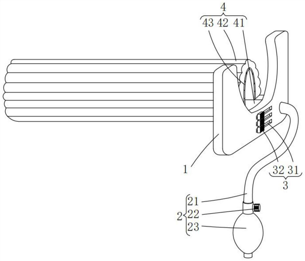
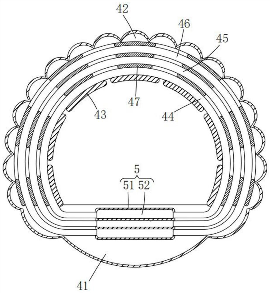
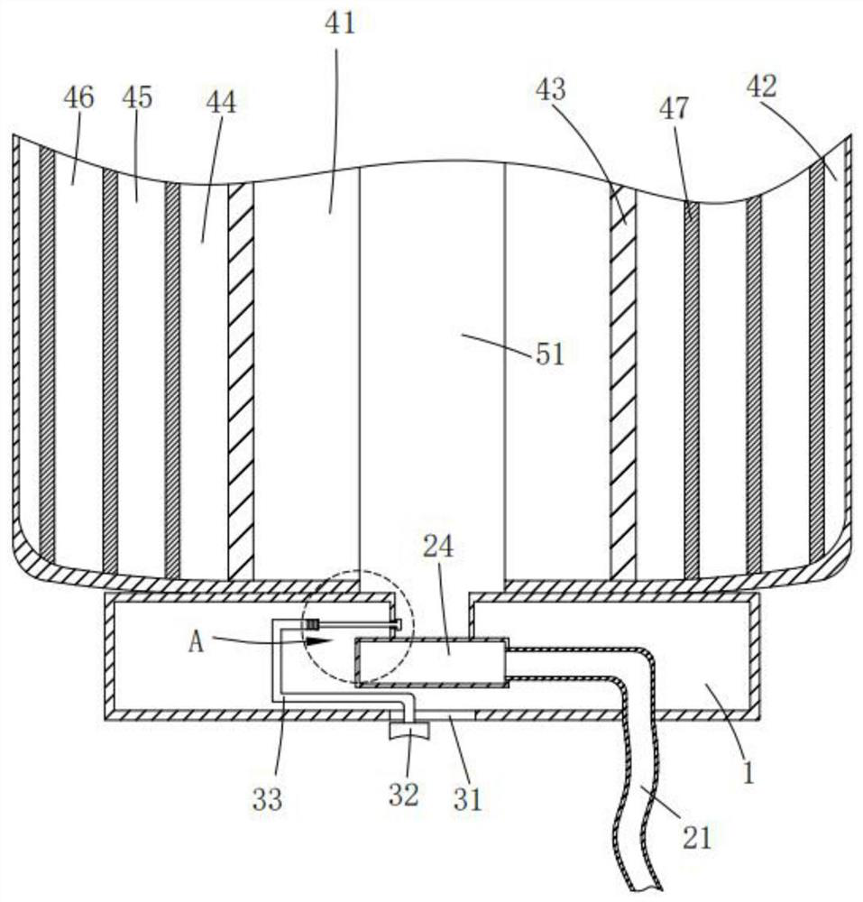
[0022] Please refer to figure 1 , figure 2 , image 3 and Figure 4 ,in, figure 1 A structural schematic diagram of a preferred embodiment of the obstetrics and gynecology clinical internal inspection device provided by the present invention; figure 2 for figure 1 Schematic cross-sectional view of the expanded structure shown; image 3 for figure 1 The schematic diagram of the cross-section of the U-shaped plate shown; Figure 4 for image 3 An enlarged schematic view of the region A shown.

[0023] Such as Figure 1-4 As shown, the obstetrics and gynecology clinical internal inspection device provided by the present invention includes: a U-shaped plate 1, an air supply structure 2, an adjustment structure 3, an expansion structure 4 and an air supply structure 5, and the air supply structure 2 and the U The front of the template 1 is conn...


PUM


Abstract

The invention provides a clinical internal examination device for gynaecology and obstetrics. The clinical internal examination device for gynaecology and obstetrics comprises a U-shaped plate, an airsupply structure, an adjusting structure, an expansion structure and an air feeding structure, wherein the air supply structure comprises an air conveying pipe, an air bag, an adjusting valve and anair cavity; the adjusting structure comprises a sliding groove, a sliding block, a connecting rod and a blocking plate; the expansion structure comprises a bottom plate, an expansion air bag, a metalsheet, a first air channel, a second air channel, a third air channel and a magnet; and the air feeding structure comprises a collecting pipe, a ventilating pipe, a telescopic groove and a sealing ring. The clinical internal examination device for the gynaecology and obstetrics has the advantages of being small in harm to a patient, easy to operate and convenient in examination.

Description

technical field [0001] The invention relates to the technical field of obstetrics and gynecology examinations, in particular to an internal clinical examination device for obstetrics and gynecology. Background technique [0002] Hysteroscopy is a new, minimally invasive gynecological diagnosis and treatment technology. It is a fiber light source endoscope used for inspection and treatment of uterine cavity, including hysteroscope, energy system, light source system, perfusion system and imaging system; It uses the front part of the mirror body to enter the uterine cavity, and has a magnifying effect on the observed parts. It is intuitive and accurate and has become the preferred inspection method for gynecological hemorrhagic diseases and intrauterine lesions. [0003] Before hysteroscopy, a dilator is required to stretch the patient's vagina. The traditional dilator is made of metal, and the surface temperature is low, which makes the patient feel uncomfortable. It is incon...

Claims


Application Information

Patent Timeline
IPC IPC(8): A61B1/32
CPCA61B1/32
Inventor 李伟贾春莲
Owner 李伟
Learn More