
MRI segmentation method based on reinforcement learning multi-scale neural network

A multi-scale segmentation and reinforcement learning technology, applied in the field of image processing, can solve the problems of inconsistent judgment severity, difference in MRI data quality, and inability to quantify, and achieve the effect of improving segmentation effect, improving segmentation accuracy, and enhancing learning.

Pending Publication Date: 2020-10-16
XIDIAN UNIV
View PDF0 Cites 0 Cited by
  • Summary
  • Abstract
  • Description
  • Claims
  • Application Information

AI Technical Summary

Problems solved by technology

[0004] Because the inflammatory area is irregular in shape, size, and distribution on the MRI data, there are problems such as inability to quantify and inconsistency in judging the severity when clinicians use MRI data to analyze patients. The difference in the quality of MRI data caused by the difference of different equipment increases the difficulty of reading images for clinicians
[0005] Existing medical image segmentation algorithms have serious missing and mis-segmented problems when segmenting inflammatory regions, which seriously affects doctors' judgment and quantitative analysis of patients, leading to wrong treatment plans. Some scholars published in Magnetic Resonance In Medicine 2019 The Measurement of synovial tissue volume in kneeosteoarthritis using a semiautomated MRI-based quantitative approach uses the prior information of the inflammatory region data and the threshold segmentation method to realize the segmentation of the inflammatory region. However, due to the lack of consideration of the multi-scale characteristics of the inflammatory region, the inflamed region poor segmentation

Method used


Image

  • MRI segmentation method based on reinforcement learning multi-scale neural network
  • MRI segmentation method based on reinforcement learning multi-scale neural network
  • MRI segmentation method based on reinforcement learning multi-scale neural network

Examples


Embodiment 1

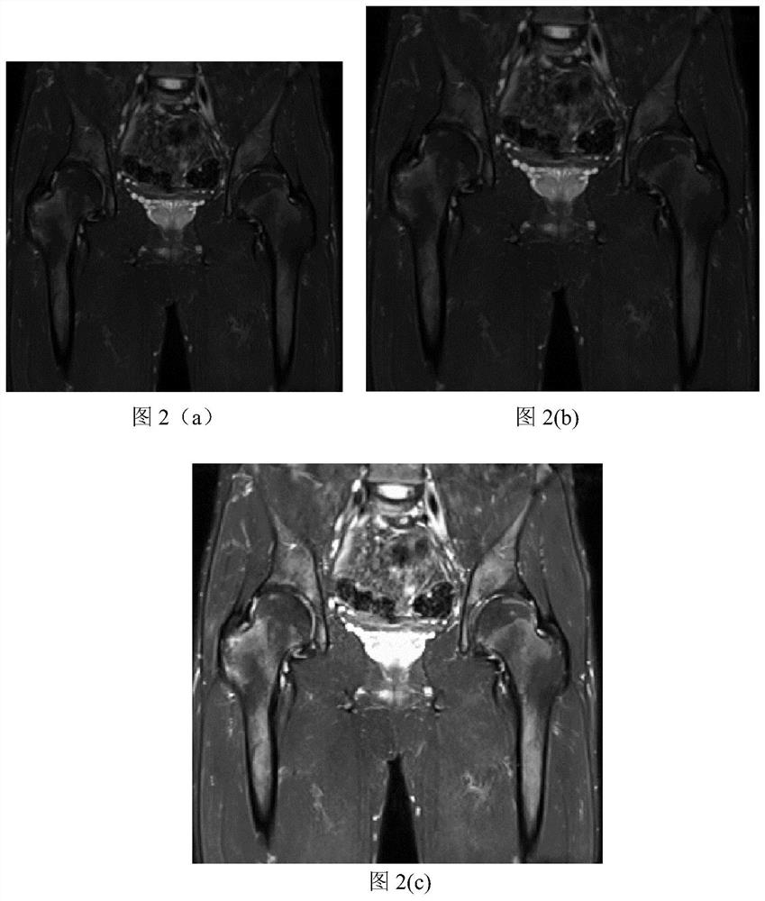
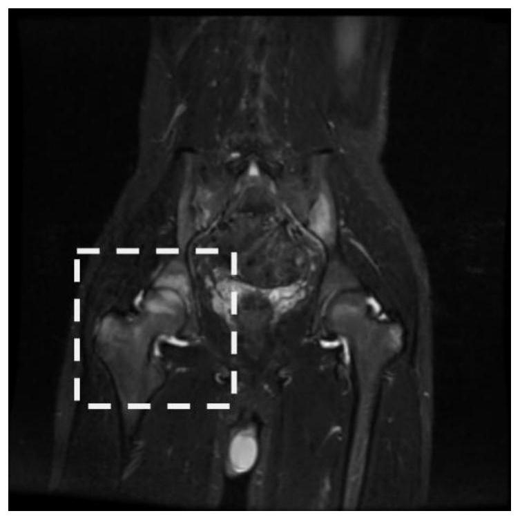
[0025] With the development of science and technology, human beings have a better understanding of spinal arthritis. More information about the inflammatory areas of spinal arthritis can be found through single-mode MRI images. In the region segmentation, the segmentation effect is often poor due to the large difference in the shape and size of the inflammatory region in the MRI image data. Aiming at the current situation, the present invention proposes an MRI segmentation method based on reinforcement learning multi-scale neural network after exploration and experimentation, which is used for segmentation of inflammatory regions in single-modality MRI images.

[0026] The invention is an MRI segmentation method based on multi-scale neural network of reinforcement learning.

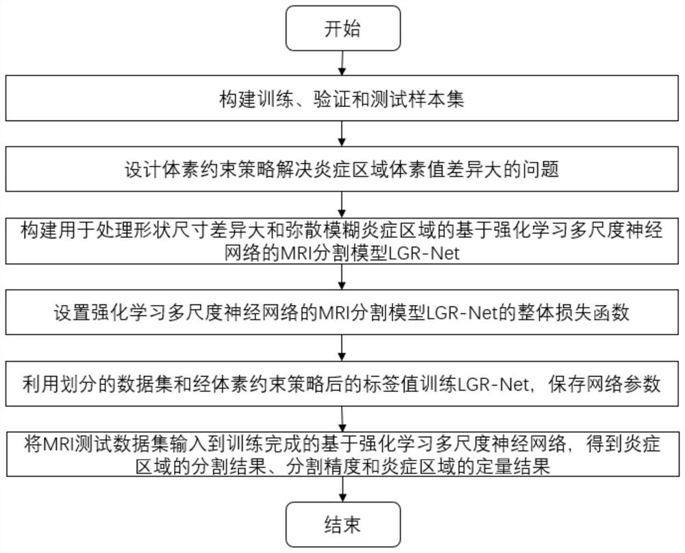
[0027] see figure 1 , including the following steps:

[0028] (1) Divide the training, verification and testing sample sets: Firstly, the original MRI data of AS patients were obtained from the hospital...

Embodiment 2

[0039] The MRI segmentation method based on reinforcement learning multi-scale neural network is the same as that in Embodiment 1, and the voxel constraint strategy described in step (2) of the present invention sets the label value of the MRI inflammation region. The voxel constraint strategy proposed by the present invention is aimed at the problem of poor segmentation effect caused by large differences in the distribution of voxel values ​​in the inflammatory region. The label value of the inflammatory region is modified according to the voxel value of the MRI data, and the label value of the inflammatory region is adaptively adjusted. According to the voxel value of the inflammatory area, it is adjusted as follows:

[0040]

[0041]

[0042] Modify the label value of the original inflammatory area according to the voxel value of the MRI data through the above formula, where y n is the original label value, y′ n is the modified label value, σ is the weighted value, p...

Embodiment 3

[0046] The MRI segmentation method based on reinforcement learning multi-scale neural network is the same as embodiment 1-2, and the construction described in the step (3) of the present invention is used to deal with the MRI based on reinforcement learning multi-scale neural network for processing large differences in shape and size and diffuse fuzzy inflammatory regions The segmentation model LGR-Net, which addresses the segmentation of multi-scale and diffusely ambiguous inflammatory regions. Including the following steps:

[0047] (3.1) Constructing a multi-scale segmentation sub-network: Firstly, aiming at the problem of large differences in the shape and size of inflammatory regions, a multi-scale convolution module GMS is constructed to extract multi-scale information. Considering the network size limitation and the commonly used convolution kernel size, the design Nine commonly used convolution kernels with different hole rates d and different sizes k are connected in ...


PUM


Abstract

The invention discloses an MRI segmentation method based on a reinforcement learning multi-scale neural network, and solves the problems of error-prone segmentation and missing segmentation of multi-scale, fuzzy and diffuse MRI inflammatory regions in the existing method. According to the method, the voxel constraint strategy of modifying the inflammatory region label value according to the voxelvalue is adopted, and the segmentation effect of the segmentation model on the inflammatory region with large voxel value difference is improved; according to the method, a multi-scale convolution module GMS is designed for inflammatory regions with large shape and size differences, and the segmentation effect of the segmentation model on the multi-scale inflammatory regions is improved; in orderto solve the problem that the segmentation model is difficult to identify the diffusion blurred inflammatory region, the reinforcement learning network is used for data enhancement, and the discrimination performance of the segmentation model for the diffusion blurred inflammatory region is improved. According to the method, wrong segmentation and missing segmentation of the multi-scale fuzzy diffusion inflammatory region are reduced, and the segmentation effect of the MRI inflammatory region is improved. The method can be used for MRI inflammatory region segmentation and quantitative analysisautomatic processing.

Description

technical field [0001] The invention belongs to the technical field of image processing, and mainly relates to the single-mode image segmentation of nuclear magnetic resonance inflammatory lesions, specifically an MRI segmentation method based on reinforcement learning multi-scale neural network, which can be used for the inflammation area in hip joint nuclear magnetic resonance image MRI Data partitioning. Background technique [0002] Ankylosing spondylitis (AS) comprises a group of related disorders characterized by inflammation of the sacroiliac joints and at sites such as the spine, peripheral joints, and tendon attachments. The etiology of AS is complex, the pathogenesis is unclear, the early clinical manifestations of patients are not typical, the traditional radiological examination is not sensitive, and the lack of specific laboratory indicators has caused great obstacles for clinicians to accurately judge AS in the early stage. In time, it will often cause serious...

Claims


Application Information

Patent Timeline
IPC IPC(8): G06T7/00G06T7/11
CPCG06T7/0012G06T7/11G06T2207/10088G06T2207/20084G06T2207/20081G06T2207/30012
Inventor 缑水平卢云飞刘宁涛曹思颖路凯童诺刘波毛莎莎焦昶哲
Owner XIDIAN UNIV
Features
  • Generate Ideas
  • Intellectual Property
  • Life Sciences
  • Materials
  • Tech Scout
  • Unparalleled Data Quality
  • Higher Quality Content
  • 60% Fewer Hallucinations
Learn More