
Lung metal marker detection device and system

A metal marking and detection device technology, applied in the field of medical devices, can solve the problems of easy diffusion of dyes, difficult to find the marked position, difficult to observe, etc., and achieves the effects of shortening operation time, reducing trauma, and being simple and convenient to use.

Pending Publication Date: 2020-11-03
SHANGHAI CHEST HOSPITAL
View PDF0 Cites 1 Cited by
  • Summary
  • Abstract
  • Description
  • Claims
  • Application Information

AI Technical Summary

Problems solved by technology

Another example is to use a dye to stain the surrounding tissue. The dye is easy to diffuse and the labeling range is enlarged, and it is difficult to observe from the outside of the lung.
For another example, by placing metal markers, the metal markers are placed inside the lungs, and the position of the markers is not easy to find during the operation

Method used


Image

  • Lung metal marker detection device and system
  • Lung metal marker detection device and system
  • Lung metal marker detection device and system

Examples


Embodiment 1

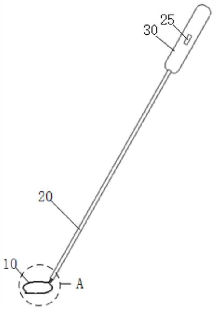
[0066] figure 1 It is a schematic structural diagram of the device in the embodiment of the present invention, such as figure 1 shown.

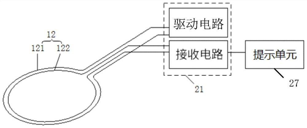
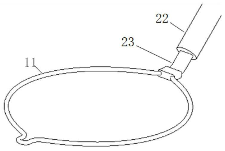
[0067] The embodiment of the present invention provides a detection device for lung metal markers, comprising: a detection head 10, a connecting rod 20 and a prompt unit 27 (the prompt unit 27 is figure 1 not shown in the figure 2 Prompt unit shown in 27). Wherein, probe head 10 comprises support frame 11 (support frame 11 is in figure 1 not shown in the image 3 The support frame 11 shown in) and the detection unit 12 arranged on the support frame 11 (the detection unit 12 is in figure 1 not shown in the figure 2 The detection unit 12 shown in ), the support frame 11 can drive the detection unit 12 to expand or collapse. The connecting rod 20 is connected with the support frame 11, and the connecting rod 20 is provided with a processing unit 21 (not shown in the figure). The detection unit 12 is coupled. The prompting unit 27 is c...

Embodiment 2

[0099] Correspondingly, an embodiment of the present invention also provides a detection system for lung metal markers, including: a marking device and a detection device for lung metal markers, wherein the detection device for lung metal markers can be obtained through embodiment 1 The detection device in is realized. The relevant technical features in Embodiment 2 and Embodiment 1 can be referred to and used for reference.

[0100] Specifically, an embodiment of the present invention also provides a detection system for lung metal markers, including: a marking device and a detection device for lung metal markers. in,

[0101] The marking device includes at least one metal marker, and the at least one metal marker is used to place inside the human body to mark the position of the lesion, and the metal marker has a certain volume.

[0102] The detection device of the lung metal marker comprises: a detection head 10, a connecting rod 20 and a prompting unit. The detection he...


PUM


Abstract

The embodiment of the invention discloses a lung metal marker detection device and system. The detection device comprises a detection head, a connecting rod and a prompt unit, wherein the detection head comprises a supporting frame and a detection unit arranged on the supporting frame, and the supporting frame can drive the detection unit to be unfolded or folded; the connecting rod is connected with the supporting frame, a processing unit is arranged on the connecting rod, and is coupled with the detection unit; the prompting unit is coupled with the processing unit; the processing unit is used for driving the detection unit to generate a detection magnetic field, receiving detection magnetic field characteristic change caused by a metal marker, detected by the detection unit, convertingthe detection magnetic field characteristic change into a prompt electric signal and sending the prompt electric signal to the prompt unit; and the prompt unit converts the received prompting electricsignal into a prompting signal so as to prompt the position of the metal marker. According to the technical scheme provided by the embodiment of the invention, the detection head can extend into a human body for use, the metal marker placed in the lung and used for positioning a focus can be quickly positioned, and the operation time is shortened when wounds of a patient are reduced.

Description

technical field [0001] The invention belongs to the technical field of medical devices, and in particular relates to a detection device and system for a lung metal marker. Background technique [0002] With the development of imaging technology and the wide application of high-resolution CT in lung examination, more and more small pulmonary nodules have been detected. Since the nodules exist in the lung and are small in size and have tissue characteristics close to normal lung tissue, it is difficult to directly observe and identify them during surgery. Effective positioning of the nodules is required to ensure accurate nodule resection. [0003] The existing nodule marking methods all have certain defects, such as placing a hook line percutaneously for marking. When placing the hook line, it is necessary to adjust the puncture multiple times and perform CT scanning, which increases the trauma of the patient. Another example is to use a dye to stain the surrounding tissue. ...

Claims


Application Information

Patent Timeline
Patent Type & Authority Applications(China)
IPC IPC(8): A61B34/20A61B17/00A61B90/00
CPCA61B17/00234A61B34/20A61B90/39A61B2034/2051A61B2034/2065A61B2090/3908A61B2090/3958A61B2090/3983
Inventor 孙加源陈军祥谢芳芳郑筱轩潘玉均
Owner SHANGHAI CHEST HOSPITAL
Learn More