Imaging method of three-dimensional blood vessel ultrasonic image and navigation equipment in ultrasonic operation

A vascular ultrasound and three-dimensional ultrasound technology, applied in the field of ultrasound, can solve the problems of difficulty in repeated acquisition, difficulty in acquiring real-time images of blood vessels, and inability to combine images in three-dimensional images, thereby achieving the effect of improving safety.

Active Publication Date: 2020-12-11
SHENZHEN DELICA MEDICAL EQUIP CO LTD
View PDF8 Cites 13 Cited by
  • Summary
  • Abstract
  • Description
  • Claims
  • Application Information

AI Technical Summary

Problems solved by technology

[0003] All of the above technologies are for the operator to hold the probe to obtain the image information of the scanning range of the ultrasonic beam at the position of the probe on the human body, and cannot obtain a larger range of three-dimensional image information beyond the position of the probe.
After the position of the probe is moved, the 3D images acquired at different positions cannot be combined into a complete image; in addition, it is also difficult to pre

Method used


Image

  • Imaging method of three-dimensional blood vessel ultrasonic image and navigation equipment in ultrasonic operation
  • Imaging method of three-dimensional blood vessel ultrasonic image and navigation equipment in ultrasonic operation
  • Imaging method of three-dimensional blood vessel ultrasonic image and navigation equipment in ultrasonic operation

Examples


Embodiment 1

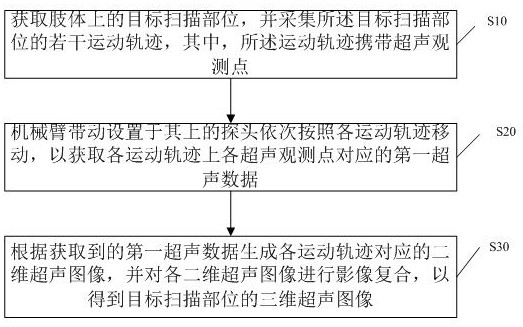
[0043] This embodiment provides an imaging method for a three-dimensional vascular ultrasound image, such as figure 1 As shown, the method includes:

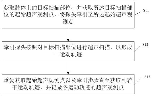
[0044] S10. Acquire a target scanning part on the limb, and collect several movement trajectories of the target scanning part, wherein the movement trajectories carry ultrasonic observation points.

[0045]Specifically, the target scanning part is a part that is preset and needs to acquire a three-dimensional image of a blood vessel, and the target scanning part is located on a limb. In addition, in order to keep the target scanning site still, after the target scanning site is determined, a preset phantom is used to fix the limbs corresponding to the target scanning site, wherein the limbs may be upper limbs or lower limbs. And after the limb is fixed by the phantom, the target scanning part is not covered by the phantom, so that the probe can contact the target scanning part and scan the target scanning part.

[0046] Exempl...

Embodiment 2

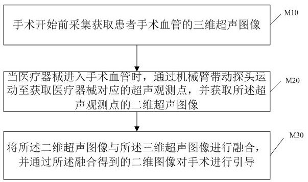
[0064] This embodiment provides a surgical guidance method based on three-dimensional ultrasound images, such as image 3 As shown, it is applied to ultrasonic equipment equipped with a mechanical arm and a probe, which includes:

[0065] M10. Acquisition and acquisition of three-dimensional ultrasound images of the patient's surgical vessels before the operation begins;

[0066] M20. When the medical device enters the surgical blood vessel, the probe is driven by the mechanical arm to move to obtain an ultrasonic observation point corresponding to the medical device, and obtain a two-dimensional ultrasonic image of the ultrasonic observation point;

[0067] M30. Fusion the two-dimensional ultrasound image and the three-dimensional ultrasound image, and guide the operation through the two-dimensional image obtained through the fusion.

[0068] Specifically, after the three-dimensional ultrasound image is acquired, when the operation starts, the robotic arm moves the probe to ...


PUM


Abstract

The invention discloses an imaging method of a three-dimensional blood vessel ultrasonic image and navigation equipment in an ultrasonic operation. The method comprises the following steps: a plurality of movement tracks at a target scanning part on a limb are determined; a mechanical arm drives a probe to move in sequence according to each movement track to obtain first ultrasonic data; and a plurality of ultrasonic images are generated according to the first ultrasonic data, and the generated ultrasonic images are subjected to image composite reconstruction to obtain a three-dimensional ultrasonic image of the target scanning part. According to the method, the mechanical arm controls the probe to move, three-dimensional image reconstruction is carried out on images collected by the probe, large-range ultrasonic image scanning and three-dimensional image reconstruction on the surface of a human body are achieved, and therefore, a complete three-dimensional ultrasonic image of large-range blood vessels in the human body is obtained, image matching and fusion can be carried out on the three-dimensional ultrasonic image and a three-dimensional DSA (or MRA and the like) image, a three-dimensional fusion image for guiding surgical operation is formed, and the vascular surgery is navigated through the three-dimensional fusion image, and the safety of the vascular surgery is improved.

Description

technical field [0001] The invention relates to the field of ultrasound technology, in particular to an imaging method of a three-dimensional blood vessel ultrasound image and a navigation device in ultrasound operation. Background technique [0002] Existing ultrasonic diagnostic equipment can be equipped with two-dimensional and three-dimensional imaging probes. Two-dimensional probes are divided into linear arrays, convex arrays, and phased arrays. They are all composed of a group of piezoelectric ceramic (or single crystal, composite material) array elements arranged on a straight line to form an array. The doctor holds the probe in different parts of the human body. scan imaging. There are two main methods for three-dimensional probes: one is mechanical scanning of three-dimensional probes, that is, a two-dimensional probe array combined with a mechanical scanning device is sealed in one probe, and the mechanical device controls the two-dimensional probe array to swing...

Claims


Application Information

Patent Timeline
IPC IPC(8): A61B8/08A61B34/10
CPCA61B8/0891A61B8/483A61B34/10A61B2034/105
Inventor 刘勇黄庆华王筱毅孟凡超熊飞严奉海丁旻昊谈家亮
Owner SHENZHEN DELICA MEDICAL EQUIP CO LTD
Why Eureka
Try Eureka