Ultrasonic navigation method and equipment for vascular operation

A technology of vascular surgery and navigation method, which is applied in the field of ultrasonic navigation method and ultrasonic navigation equipment for vascular surgery, which can solve the problems of repeated acquisition difficulties, affecting the accuracy of ultrasound equipment in vascular surgery navigation of extremities, and the inability to navigate vascular surgery of extremities, etc., to achieve The effect of improving safety

Active Publication Date: 2020-12-11
SHENZHEN DELICA MEDICAL EQUIP CO LTD
View PDF15 Cites 3 Cited by
  • Summary
  • Abstract
  • Description
  • Claims
  • Application Information

AI Technical Summary

Problems solved by technology

[0003] All of the above technologies are for the operator to hold the probe to obtain the image information of the scanning range of the ultrasonic beam at the position of the probe on the human body, and cannot obtain a larger range of three-dimensional image information beyond the position of the probe.
After the position of the probe is moved, the 3D images acquired at different positions cannot be combined into a complete image; in addition, it is also difficult to precisely position the probe at the same position for multiple repeated acquisitions
Especially for the long arteries or veins on the limbs, it is difficult to collect or repeat the complete real-time images of the blood vessels, which affects the navigation accuracy of the ultrasound equipment in the vascular surgery of the limbs, making it impossible for the ultrasound equipment to navigate the vascular surgery of the limbs

Method used


Image

  • Ultrasonic navigation method and equipment for vascular operation
  • Ultrasonic navigation method and equipment for vascular operation
  • Ultrasonic navigation method and equipment for vascular operation

Examples


Embodiment 1

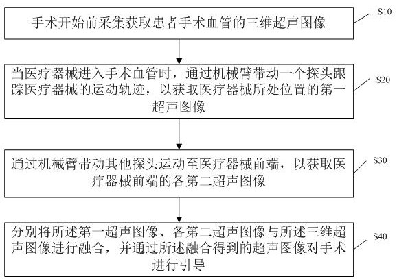
[0045] This embodiment provides an ultrasonic navigation method for vascular surgery, such as figure 1 As shown, the method includes:

[0046] S10. Acquiring three-dimensional ultrasound images of the surgical blood vessels of the patient before the operation begins;

[0047] S20. When the medical device enters the surgical blood vessel, the robotic arm drives a probe to track the movement trajectory of the medical device, so as to obtain the first ultrasonic image of the position of the medical device;

[0048]S30. Drive other probes to move to the front end of the medical device through the mechanical arm, so as to obtain the second ultrasonic images of the front end of the medical device;

[0049] S40. Fusion the first ultrasound image, each second ultrasound image, and the three-dimensional ultrasound image respectively, and use the fusion ultrasound image to guide the operation.

[0050] In this embodiment, the ultrasonic image of the front end of the medical device and...

Embodiment 2

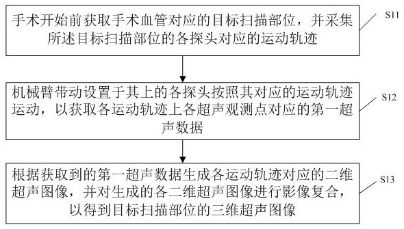
[0076] This embodiment provides an ultrasonic navigation method for vascular surgery, such as Figure 4 As shown, the application is configured with an ultrasonic device with a robotic arm and at least two probes, which includes:

[0077] M10. Acquiring three-dimensional ultrasound images and three-dimensional images of the patient's surgical vessels before the operation starts, wherein the three-dimensional images are one of DSA images, MRA images and CTA images;

[0078] M20. Perform image registration on the three-dimensional ultrasound image and the three-dimensional image to obtain a three-dimensional fusion image of the surgical vessel;

[0079] M30. When the medical device enters the surgical blood vessel, a probe is driven by the mechanical arm to track the movement trajectory of the medical device, so as to obtain the first ultrasonic image of the position of the medical device;

[0080] M40. Drive other probes to move according to their corresponding motion trajecto...


PUM


Abstract

The invention discloses an ultrasonic navigation method and equipment for a vascular operation. The method comprises the steps that one probe is used for tracking the movement track of a medical instrument to obtain a first ultrasonic image of the position where the medical instrument is located; second ultrasonic images of the front end of the medical instrument are obtained through the movementof the other probes according to the corresponding movement tracks; and the first ultrasonic image, the second ultrasonic images and a three-dimensional ultrasonic image are fused, and the operation is guided through a two-dimensional image obtained by fusion. According to the method, the ultrasonic images of the front end of the medical instrument and the ultrasonic image of the position where the medical instrument is located are obtained in the operation process, the obtained ultrasonic images and the three-dimensional ultrasonic image are fused, the blood flow information of the medical instrument is determined according to the image obtained through fusion, and the vascular operation is navigated according to the blood flow information, so that application of the ultrasonic equipmentin limb blood vessel navigation is realized, and meanwhile, the safety of the vascular operation is improved.

Description

technical field [0001] The invention relates to the field of ultrasonic technology, in particular to an ultrasonic navigation method for vascular surgery and an ultrasonic navigation device. Background technique [0002] Existing ultrasonic diagnostic equipment can be equipped with two-dimensional and three-dimensional imaging probes. Two-dimensional probes are divided into linear arrays, convex arrays, and phased arrays. They are all composed of a group of piezoelectric ceramic (or single crystal, composite material) array elements arranged on a straight line to form an array. The doctor holds the probe in different parts of the human body. scan imaging. There are two main methods for three-dimensional probes: one is mechanical scanning of three-dimensional probes, that is, a two-dimensional probe array combined with a mechanical scanning device is sealed in one probe, and the mechanical device controls the two-dimensional probe array to swing back and forth to obtain two-...

Claims


Application Information

Patent Timeline
Patent Type & Authority Applications(China)
IPC IPC(8): A61B34/20
CPCA61B34/20A61B2034/2063
Inventor 华扬刘玉梅王筱毅黄庆华周赤宜孟凡超严奉海
Owner SHENZHEN DELICA MEDICAL EQUIP CO LTD
Why Eureka
Try Eureka