3D printed tooth root stent for alveolar bone site preservation, and preparation method thereof

A 3D printing and alveolar bone technology, applied in the field of medical stents, can solve the problems of inability to effectively prevent the absorption of alveolar ridges and promote new bone, achieve good biological characteristics and promote the formation of

Pending Publication Date: 2020-12-11
STOMATOLOGY AFFILIATED STOMATOLOGY HOSPITAL OF GUANGZHOU MEDICAL UNIV
View PDF5 Cites 1 Cited by
  • Summary
  • Abstract
  • Description
  • Claims
  • Application Information

AI Technical Summary

Problems solved by technology

[0007] In order to overcome the defect that the bone substitute material and / or biofilm used for the preservation of the alveolar bone site described in the above prior art cannot effectively prevent the absorption of the alveolar ridge and cannot promote the formation of new bone, the present invention provides a The 3D printed tooth root bracket preserved in the alveolar bone site. When the 3D printed tooth root bracket is used for the preservation of the alveolar bone site, it can effectively preserve the horizontal width of the alveolar ridge and the vertical height of the buccal bone plate of the alveolar ridge after tooth extraction. , and promote the formation of new bone in the alveolar socket

Method used


Image

  • 3D printed tooth root stent for alveolar bone site preservation, and preparation method thereof
  • 3D printed tooth root stent for alveolar bone site preservation, and preparation method thereof
  • 3D printed tooth root stent for alveolar bone site preservation, and preparation method thereof

Examples


Embodiment 1

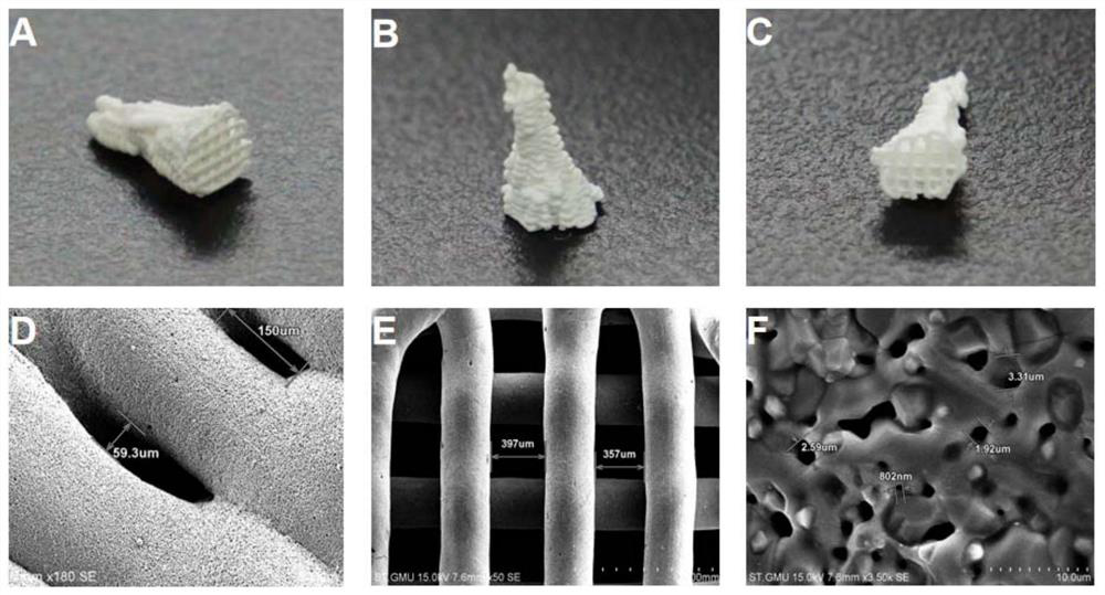
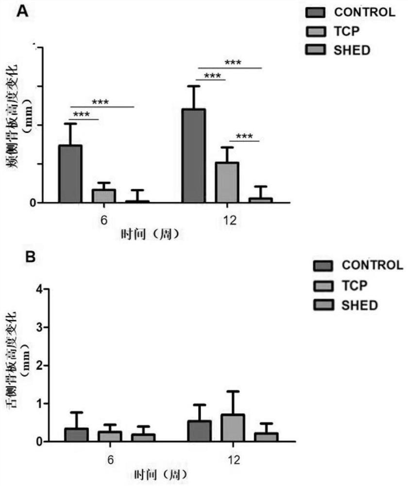
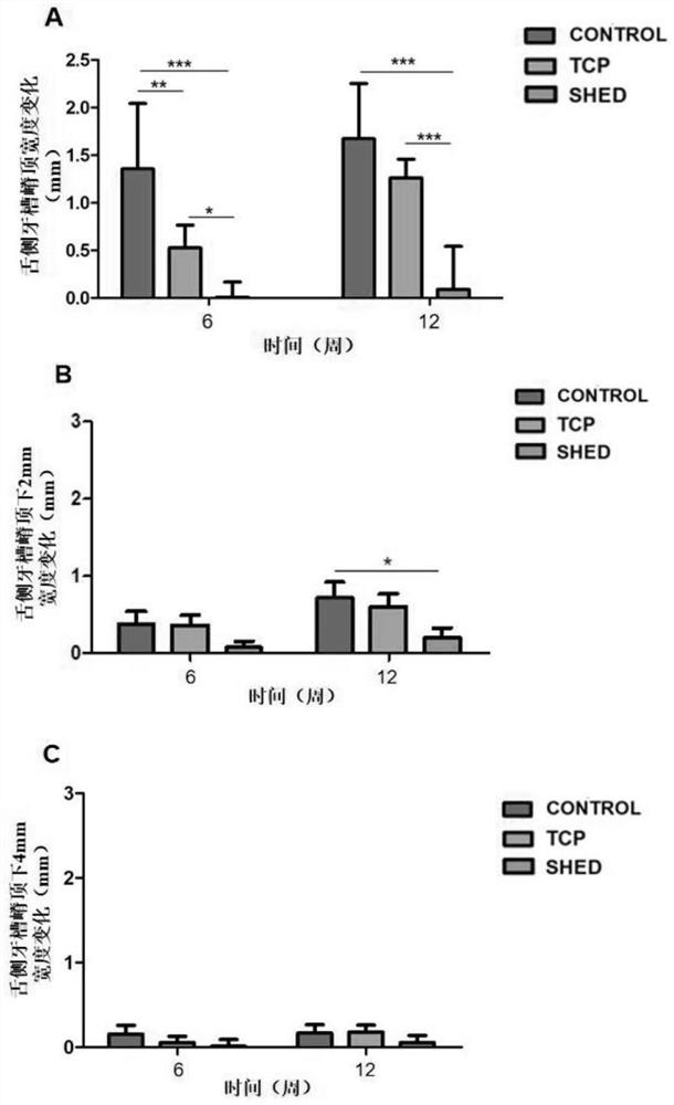
[0037] This embodiment provides a 3D printed tooth root bracket for the preservation of alveolar bone sites. The 3D printed tooth root bracket is a hollow three-dimensional connected structure, the main material is β-tricalcium phosphate, and deciduous tooth pulp stem cells are adhered on the surface and processed. Osteogenic induction.

[0038] The preparation method of the 3D printing tooth root bracket of the present embodiment is as follows:

[0039]S1. Mandible data were collected by cone beam projection computerized tomography (corn bean computed tomography, CBCT). The scanning layer thickness was 0.25mm. After the scanning was completed, the jaw shape was reconstructed, and the standard dicom format file was saved. Import the data into MIMICS 17.0, adjust the threshold of the image, and optimize to make the image clear and complete. Output the processed image data in STL format, continue to optimize, carefully remove the tissues other than P2-P4 of the mandible, and ob...


PUM

PropertyMeasurementUnit
porosityaaaaaaaaaa
porosityaaaaaaaaaa
survival rateaaaaaaaaaa

Abstract

The invention discloses a 3D printed tooth root stent for alveolar bone site preservation, and a preparation method thereof. The 3D printed tooth root stent for alveolar bone site preservation is of ahollow three-dimensional communication structure; a main body material is beta-tricalcium phosphate; stem cells from human exfoliated deciduous teeth are adhered to the surface; and osteogenic induction is carried out. According to the invention, advantages of a biological ceramic material (beta-tricalcium phosphate), the stem cells and a 3D printing technology are combined and jointly applied tosite preservation; the 3D printed TCP stent has good biological characteristics; furthermore, the stem cells from human exfoliated deciduous teeth can serve as good bone tissue engineering seed cellsloaded on the stent; the stem cells from human exfoliated deciduous teeth are combined with the TCP stent material; therefore, the horizontal width of an alveolar bone and the vertical height of an alveolar ridge buccal bone plate after tooth extraction can be effectively preserved; and the formation of new bones in alveolar fossa can be promoted.

Description

technical field [0001] The invention relates to the technical field of medical brackets, and more specifically, to a 3D printed tooth root bracket for alveolar bone site preservation and a preparation method thereof. Background technique [0002] Clinically, factors such as periodontal disease, periapical disease and trauma often lead to tooth loss. The shape and volume of the alveolar bone are determined by the storage and loss, shape, growth direction and long axis of the teeth. When the teeth are lost or extracted, the physiological process of hard tissue and soft tissue remodeling will lead to changes in the height and width of the alveolar ridge. The degree of reduction was related to several variables, including alveolar socket size, mucosal thickness, metabolic factors, and functional load. Studies have shown that the horizontal bone loss rate is 29% to 63% and the vertical bone loss rate is 11% to 22% 6 months after tooth extraction. The resorption pattern is chara...

Claims


Application Information

Patent Timeline
Patent Type & Authority Applications(China)
IPC IPC(8): A61F2/28
CPCA61F2/28A61F2002/2817A61F2002/2839A61F2002/2889A61F2002/30985
Inventor 周苗葛立宏曾素娟李云阳谌斯李树祎耿远明
Owner STOMATOLOGY AFFILIATED STOMATOLOGY HOSPITAL OF GUANGZHOU MEDICAL UNIV
Why Eureka
Try Eureka