CT-based three-dimensional jawbone image segmentation modeling method and device and terminal equipment

A CT image and image segmentation technology, applied in the field of image models, can solve problems such as poor segmentation accuracy, and achieve the effects of accurate judgment, improved stability and response efficiency

Inactive Publication Date: 2020-12-29
北京羽医甘蓝信息技术有限公司 +1
View PDF6 Cites 2 Cited by
  • Summary
  • Abstract
  • Description
  • Claims
  • Application Information

AI Technical Summary

Problems solved by technology

[0004] In the related technologies of CT-based maxillofacial fracture diagnosis, upper and lower jaw segmentation and three-dimensional visualization are of great significance for facial surgery diagnosis. At present, the main method is semi-automatic way (usually threshold method or region growing method) to segment the upper and lower jaws, the segmentation accuracy of the existing software is poor

Method used


Image

  • CT-based three-dimensional jawbone image segmentation modeling method and device and terminal equipment
  • CT-based three-dimensional jawbone image segmentation modeling method and device and terminal equipment
  • CT-based three-dimensional jawbone image segmentation modeling method and device and terminal equipment

Examples


Embodiment Construction

[0023] It should be noted that, in the case of no conflict, the embodiments and features in the embodiments of the present invention can be combined with each other.

[0024] Specific embodiments of the present invention will be described in detail below in conjunction with the accompanying drawings. It should be understood that the specific embodiments described here are only used to illustrate and explain the present invention, and are not intended to limit the present invention.

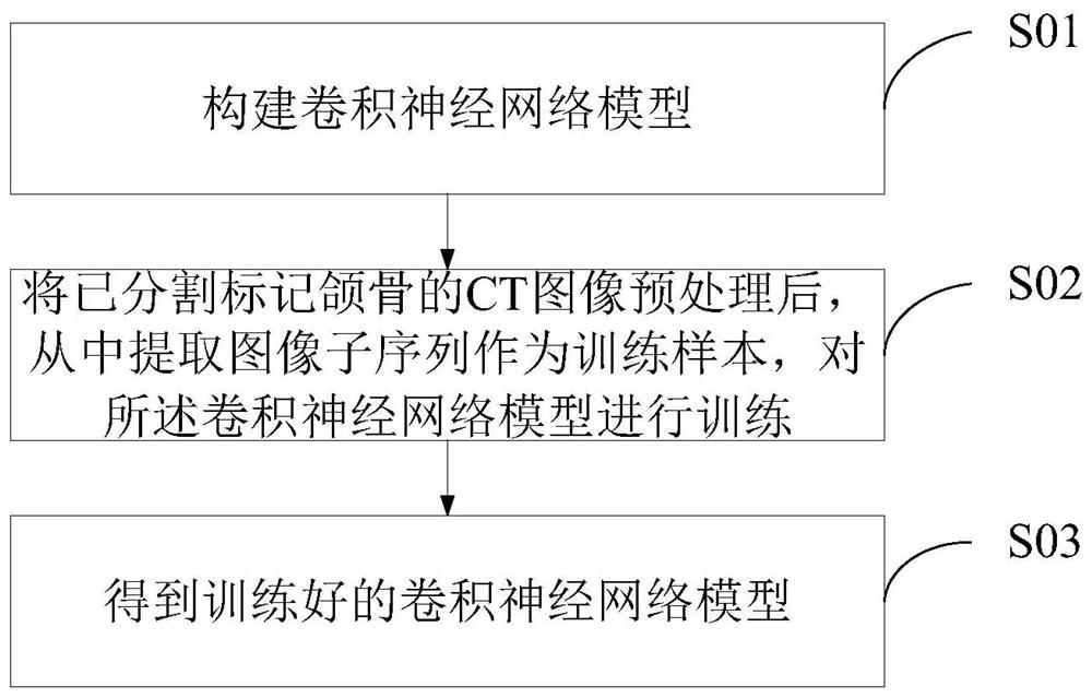
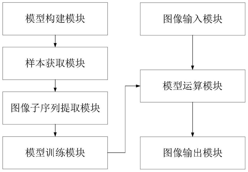
[0025] figure 1 It is a schematic flow chart of a CT-based three-dimensional jaw image segmentation and modeling method provided by an embodiment of the present invention, such as figure 1 shown. In one embodiment of the present invention, a CT-based three-dimensional jaw image segmentation modeling method is provided, the method comprising: constructing a convolutional neural network model; preprocessing the CT image of the segmented and marked jaw , extract image subsequence therefrom as trai...


PUM


Abstract

The invention relates to the field of image models, and provides a three-dimensional jawbone image segmentation modeling method based on CT, and the method comprises the steps of constructing a convolutional neural network model; preprocessing the CT image with the segmented and marked jawbone, extracting an image sub-sequence from the CT image as a training sample, and training the convolutionalneural network model to obtain a trained convolutional neural network model, wherein the trained convolutional neural network model is used for obtaining an image sub-sequence extracted from a CT image to be segmented, and obtaining a segmentation result corresponding to the image sub-sequence after processing, wherein the segmentation result is used for obtaining a three-dimensional jaw bone segmentation result of the CT image to be segmented after integration. Meanwhile, the invention further provides a corresponding three-dimensional jaw image segmentation modeling device based on CT, and terminal equipment. The embodiment provided by the invention is suitable for establishing a segmentation model for extracting the three-dimensional jawbone region from the CT image.

Description

technical field [0001] The present invention relates to the field of image models, in particular to a CT-based three-dimensional jaw image segmentation modeling method, a CT-based three-dimensional jaw image segmentation modeling device, a terminal device and a corresponding storage medium. Background technique [0002] As a common fracture with complex structure, maxillofacial fractures are difficult to diagnose only by clinical symptoms and signs, and often require auxiliary diagnosis of imaging. Compared with traditional X-ray films, the three-dimensional images of maxillofacial CT can more clearly and intuitively show the location of fractures and the direction of displacement, and are considered the "gold standard" for the diagnosis of maxillofacial fractures. However, due to the relatively large information contained in CT images, the diagnosis and treatment model based on this method relies on personnel who are proficient in imaging knowledge and professional equipmen...

Claims


Application Information

Patent Timeline
Patent Type & Authority Applications(China)
IPC IPC(8): G06T7/10G06N3/04G06N3/08G16H50/20
CPCG06T7/10G06N3/08G16H50/20G06T2207/30008G06T2207/10081G06N3/045
Inventor 徐子能贺洋揭璧朦张益仝雁行彭歆丁鹏白海龙
Owner 北京羽医甘蓝信息技术有限公司
Why Eureka
Try Eureka