Fatty liver intelligent grading evaluation method based on abdominal CT

A technique for fatty liver and abdomen, which is applied in the field of intelligent grading and assessment of fatty liver based on abdominal CT, which can solve the problems of time-consuming and labor-intensive

Pending Publication Date: 2021-01-05
YANCHENG INST OF TECH
View PDF4 Cites 2 Cited by
  • Summary
  • Abstract
  • Description
  • Claims
  • Application Information

AI Technical Summary

Problems solved by technology

Manual segmentation of CT images is time-consuming and labor-intensive, and then a semi-automatic segmentation method appeared, but it

Method used


Image

  • Fatty liver intelligent grading evaluation method based on abdominal CT
  • Fatty liver intelligent grading evaluation method based on abdominal CT
  • Fatty liver intelligent grading evaluation method based on abdominal CT

Examples


Embodiment 1

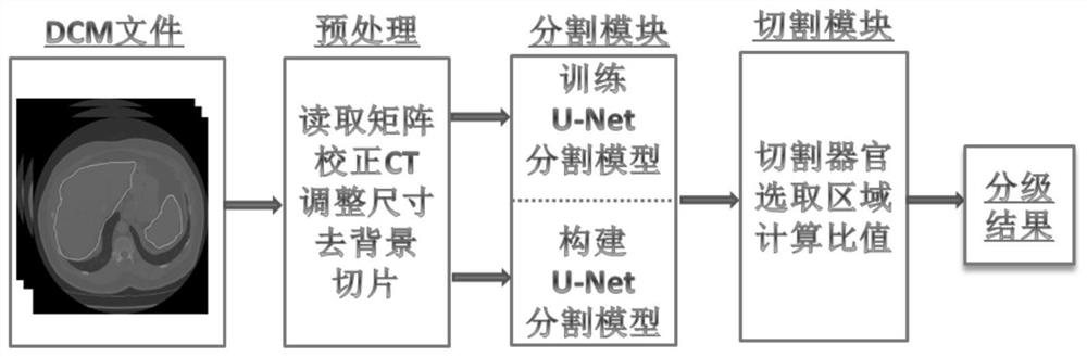
[0045] The fatty liver intelligent grading evaluation method and system based on abdominal CT in one embodiment of the present invention specifically includes the following steps:

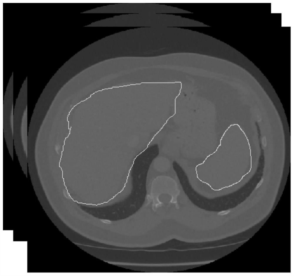
[0046] Step 1, for example figure 2The preprocessing of the patient’s abdominal CT image data is as follows: use the SimpleITK toolkit to read the DCM image file, use the Numpy toolkit to convert the file into an array matrix, and use the window width and window level technology to correct the CT value of the slice, the window width value is 400hu, The window level value is 100hu, and the bilinear interpolation technology is used to adjust the size of the CT slice. The adjusted slice size is 256*256, and the image connected area method is used to eliminate the equipment background interference information in the image;

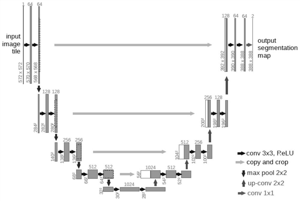
[0047] Step 2. Input the CT slice image, use the U-Net segmentation model to segment the CT slice of the patient, output the respective mask maps of the liver tissue and spleen ti...


PUM


Abstract

The invention relates to a fatty liver intelligent grading evaluation method based on abdominal CT, and relates to the field of intelligent medical image diagnosis. The method comprises the followingsteps: reading an abdominal CT image of a patient, selecting nearby four layers of slices corresponding to the maximum area of liver and spleen to construct a training sample set, and carrying out thepreprocessing of the data of the training sample set; constructing a UNet segmentation network model, sending the training sample to the segmentation network model for supervised learning, and aftertraining convergence of the segmentation model, using the model to segment CT slices to segment liver tissues and spleen tissues in the slices; respectively carrying out gridding cutting on liver tissues and spleen tissues to obtain a plurality of small rectangular areas with the same area, randomly selecting five small rectangular areas in the two tissues as sampling areas, calculating respectivegray average values as a liver CT value and a spleen CT value, and finally grading the fatty liver according to a liver/spleen CT ratio. According to the method, full-automatic segmentation of the liver tissue and the spleen tissue based on the abdominal CT image is realized, so that the fatty liver is intelligently graded and evaluated.

Description

technical field [0001] The invention relates to the field of intelligent medical image diagnosis, in particular to an intelligent grading and evaluation method for fatty liver based on abdominal CT. Background technique [0002] Today's human diet structure and activity patterns have undergone tremendous changes. A well-off life allows people to obtain excess fat in their diet, but physical labor is much reduced, which indirectly induces many diseases of affluence. Fatty liver, as a relatively common digestive system disease, is caused by abnormal fat metabolism in liver cells due to various reasons, resulting in excessive accumulation of fat. Early diagnosis and timely treatment of fatty liver can often return to normal. Clinically, B-ultrasound, CT and other imaging techniques are often used for qualitative and quantitative diagnosis of fatty liver. Due to the accumulation of fat in CT examination, fatty liver is mainly manifested as a decrease in liver density. In imagin...

Claims


Application Information

Patent Timeline
IPC IPC(8): G06T7/11G06T7/00G06N3/08G06N3/04A61B6/03A61B6/00
CPCG06T7/0012G06T7/11G06N3/08A61B6/5217A61B6/032G06T2207/10081G06T2207/30056G06N3/045
Inventor 王东洋
Owner YANCHENG INST OF TECH
Why Eureka
Try Eureka