Augmented reality module for microscope

An augmented reality and microscope technology, applied in microscopes, instruments, optics, etc., can solve the inconvenience of microscopes and other problems, and achieve the effects of saving expensive expenses, accelerating development, and small size

Pending Publication Date: 2021-02-09
HUAZHONG UNIV OF SCI & TECH
View PDF0 Cites 3 Cited by
  • Summary
  • Abstract
  • Description
  • Claims
  • Application Information

AI Technical Summary

Problems solved by technology

[0005] Aiming at the defects of the prior art, the purpose of the present invention is to provide an augmented reality module for a microscope, which aims to solve the inconvenience of traditional microscopes in imaging

Method used


Image

  • Augmented reality module for microscope
  • Augmented reality module for microscope
  • Augmented reality module for microscope

Examples


Embodiment

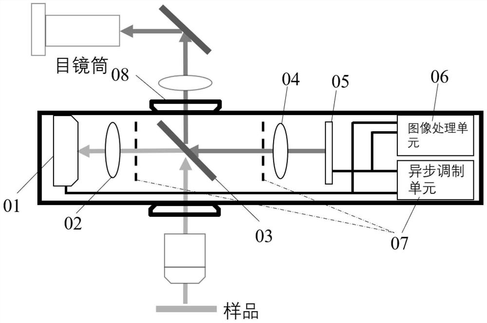
[0027] This embodiment provides an augmented reality module for a microscope. The principle of the whole scheme is as follows figure 1 As shown, it includes a camera 01 , a first imaging lens 02 , a beam splitter 03 , a second imaging lens 04 , a display screen 05 , an image processing unit 06 , an asynchronous modulation unit 07 , and a housing 08 .

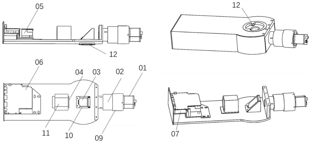
[0028] figure 2 What is shown is a schematic structural diagram of an embodiment of an augmented reality module. figure 2 The shell and the microscope interface are made of aluminum alloy, and the shell is designed with a dovetail groove 12 that matches the mainstream microscope interface. A second imaging lens holder 11 and a beam splitter holder 10 are designed inside the housing. Wherein, the second imaging lens holder 11 is designed with a fine-tuning structure, which can adjust the position of the lens relative to the beam splitter.

[0029] In this embodiment, camera 01 uses a camera with a resolution of 1920×1200 and ...


PUM


Abstract

The invention discloses an augmented reality module for a microscope, and belongs to the field of biomedical instrument design. The augmented reality module comprises a camera, a first imaging lens, abeam splitter, a second imaging lens, a display screen, an image processing unit and an asynchronous modulation unit. A sample image is divided into two paths by the beam splitter, and one path is directly coupled into a microscope visual light path; the other path is converted into an electric signal through a camera after passing through a first imaging lens, the electric signal is input into an image processing unit, the image processing unit analyzes and recognizes the electric signal and then images the image on a display screen, and the display screen image is divided into two paths bythe same beam splitter after passing through a second imaging lens; and one path is prevented from imaging on a camera target surface through a display screen and an asynchronous modulation unit, andthe other path is coupled into a microscope visual light path. According to the augmented reality module provided by the invention, a traditional existing microscope can be directly modified into an intelligent microscope capable of being used for auxiliary diagnosis, and a computer-aided diagnosis system can be accessed under the condition of not changing the use habits of doctors.

Description

technical field [0001] The invention belongs to the field of biomedical instrument design, and more specifically relates to an augmented reality module for a microscope. Background technique [0002] Pathological examination plays an irreplaceable role in the screening and treatment of tumors and cancers. In recent years, the incidence of clinical cancer and tumors has been high, and the demand for pathological examinations has also increased. Pathological examination is a pathomorphological method used to examine pathological changes in body organs, tissues or cells. In order to explore the disease process of organs, tissues or cells, some pathological and morphological examination method can be adopted to check the lesions they have, discuss the causes of lesions, pathogenesis, and the development process of lesions, and finally make a pathological diagnosis. diagnosis. The examination method of pathological morphology usually needs to cut out a lesion tissue of a certai...

Claims


Application Information

Patent Timeline
Patent Type & Authority Applications(China)
IPC IPC(8): G02B21/36G02B21/00
CPCG02B21/0012G02B21/361G02B21/367
Inventor 李宁刘晓翔蒋国庆吕晓华曾绍群
Owner HUAZHONG UNIV OF SCI & TECH
Why Eureka
Try Eureka