3D medical image segmentation method, device and storage medium based on layered perceptual fusion

A medical image and 3D technology, which is applied in image analysis, image enhancement, image data processing, etc., can solve the problems that a single model cannot make full use of prediction, inaccurate, etc., and achieve the effect of efficient and accurate 3D medical image segmentation

Active Publication Date: 2021-09-03
GUANGZHOU YUNRUNDA DATA SERVICES CO LTD
View PDF5 Cites 0 Cited by
  • Summary
  • Abstract
  • Description
  • Claims
  • Application Information

AI Technical Summary

Problems solved by technology

[0006] In order to overcome the deficiencies in the above-mentioned prior art, the object of the present invention is to provide a 3D medical image segmentation method, device and storage medium based on layered perceptual fusion, so as to divide the 3D medical image into H, W, and C directions. Three 2D images and several small 3D images, using the fusion of the 2D channel sequence relationship model and the 3D model, solve the problem that a single model cannot be fully utilized and the prediction is inaccurate, and a voting mechanism is established based on the fusion of multiple models to achieve efficient The purpose of accurate 3D medical image segmentation

Method used


Image

  • 3D medical image segmentation method, device and storage medium based on layered perceptual fusion
  • 3D medical image segmentation method, device and storage medium based on layered perceptual fusion
  • 3D medical image segmentation method, device and storage medium based on layered perceptual fusion

Examples


Embodiment

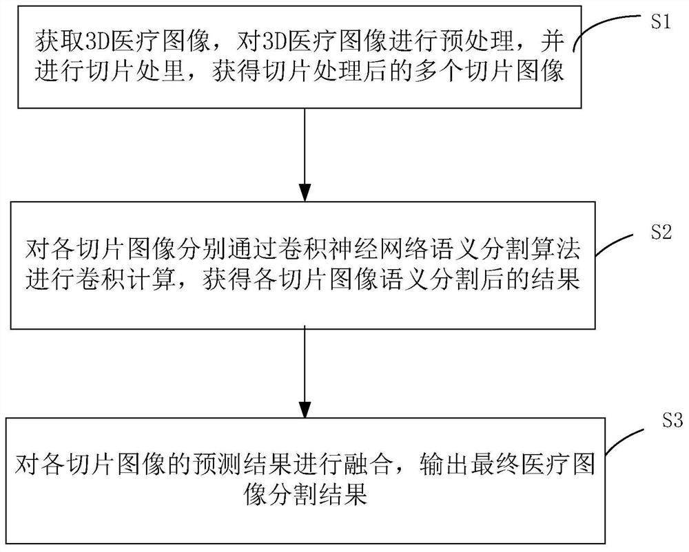
[0082] Figure 5 It is a flow chart of 3D medical image segmentation based on layered perceptual fusion in an embodiment of the present invention. In an embodiment of the present invention, a 3D medical image segmentation method based on layered perceptual fusion includes:

[0083] Step 1, data creation, slicing, and enhancement

[0084] Step 1.1: use marking software to mark the 3D medical image, and divide it into two parts: the target and the background. The target represents the target area, that is, the organ pathological tissue image area, and the background represents the non-organ part. If there are multiple pathological tissues, different pixels can be used to distinguish them, for example, pixel 1 is the tumor tissue, pixel 2 is the periphery of the tumor, and so on.

[0085] Step 1.2, after the marking is completed, check the correctness of the data. For example, use programming to check whether the data distribution of the background and lesion area is backgroun...


PUM


Abstract

The invention discloses a 3D medical image segmentation method, device and storage medium based on layered perceptual fusion. The method includes the following steps: step S1, obtaining a 3D medical image for preprocessing, and preprocessing the preprocessed 3D medical image Carry out slices to obtain multiple sliced ​​images; step S2, perform convolution calculation on each sliced ​​image respectively through the convolutional neural network semantic segmentation algorithm, and obtain the result of each sliced ​​image after semantic segmentation; step S3, predict the result of each sliced ​​image Fusion is performed to output the final medical image segmentation results.

Description

technical field [0001] The present invention relates to the technical field of image segmentation, in particular to a 3D medical image segmentation method, device and storage medium based on layered perceptual fusion. Background technique [0002] The object of medical image processing is a variety of medical images with different imaging mechanisms. The types of medical imaging widely used in clinical practice mainly include X-ray imaging (X-CT), nuclear magnetic resonance imaging (MRI), nuclear medical imaging (NMI) and ultrasonic imaging. (UI) four categories. In the current imaging medical diagnosis, it is mainly to find the lesion by observing a group of two-dimensional slice images, which often needs to be judged by the doctor's experience. Using computer image processing technology to analyze and process two-dimensional slice images, realize the segmentation and extraction, three-dimensional reconstruction and three-dimensional display of human organs, soft tissues a...

Claims


Application Information

Patent Timeline
Patent Type & Authority Patents(China)
IPC IPC(8): G06T7/00G06T7/11G06N3/04
CPCG06T7/0012G06T7/11G06T2207/10012G06T2207/20084G06T2207/20021G06T2207/30096G06N3/045
Inventor 孟令龙
Owner GUANGZHOU YUNRUNDA DATA SERVICES CO LTD
Why Eureka
Try Eureka