Semi-supervised medical image segmentation method based on geometric consistency constraint

A medical image and image segmentation technology, applied in image analysis, neural learning methods, image data processing, etc., can solve the problems of not considering the difficulty of segmentation in different regions, not considering the quality of the original image data, affecting the segmentation performance, etc. , to achieve the effect of improving training efficiency, good generalization, and reducing uncertainty

Pending Publication Date: 2022-08-05
ZHEJIANG UNIV
View PDF0 Cites 0 Cited by
  • Summary
  • Abstract
  • Description
  • Claims
  • Application Information

AI Technical Summary

Problems solved by technology

However, these methods ignore two difficulties in medical image segmentation
One is that it does not consider the different degrees of difficulty of different regions, resulting in unclear segmentation of difficult regions such as target boundaries
The second is that the uneven quality of the original image data is not considered, resulting in high uncertainty prediction results, which in turn affects the segmentation performance
Therefore, how to effectively use the information differences in different regions of the image to obtain a low-uncertainty, high-precision medical image segmentation model is a huge challenge.

Method used


Image

  • Semi-supervised medical image segmentation method based on geometric consistency constraint
  • Semi-supervised medical image segmentation method based on geometric consistency constraint
  • Semi-supervised medical image segmentation method based on geometric consistency constraint

Examples


Embodiment Construction

[0042] The present invention will be described in further detail below in conjunction with the accompanying drawings and specific examples.

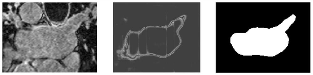
[0043] Medical image segmentation is an important part in the diagnosis and analysis of many diseases. In recent years, with the rapid development of imaging technology, the number of medical images has increased exponentially. The study of efficient and accurate automatic segmentation algorithms for medical images is of great significance for promoting the development of medical research and human health. However, the data quality of medical images is often uneven and the target boundary information is complex, such as figure 1 As shown, the usual segmentation results have high uncertainty. The present invention fuses the geometric structure information of the segmentation target, and achieves better results in the accuracy and completeness of the segmentation. The following takes the public dataset of the 2018 MICCAI Left Atrial Segm...


PUM


Abstract

The invention discloses a semi-supervised medical image segmentation method based on geometric consistency constraint. In order to solve the problems of high cost, time consumption and labor consumption of medical image annotation acquisition, the model based on geometric consistency constraint and dual deep neural network is constructed, and accurate automatic segmentation of the medical image can be realized only by a small number of annotated images. In addition, the method fully considers the characteristics of large noise, boundary segmentation blur and the like of a medical image, and utilizes an auxiliary task to learn geometric structure information of a segmentation target, thereby helping the model to better realize segmentation of a boundary blur region. And meanwhile, target segmentation is carried out from different perspectives by using the dual deep neural network model, so that the uncertainty of segmentation is reduced, and the prediction accuracy is improved. The method can be suitable for various medical image segmentation tasks, and meanwhile, the segmentation precision of the method is remarkably improved compared with that of an existing advanced algorithm. The method can effectively relieve the workload of imageologists.

Description

technical field [0001] The invention belongs to the field of automatic image segmentation in medical image analysis, and is particularly aimed at intelligent image segmentation in the case of insufficient labeled image data and sufficient unlabeled data. Background technique [0002] In practical clinical applications, accurate and robust segmentation of organs or diseased regions based on medical images plays a very important role. Accurate segmentation results can help doctors to better diagnose and quantitatively analyze diseases and provide theoretical basis for the next diagnosis and treatment plan. In recent years, with the rapid development of the medical industry and the continuous advancement of medical imaging technology, medical image data has exploded. However, medical image analysis requires a lot of expert knowledge and time consumption. Therefore, the realization of automatic segmentation of medical images can effectively relieve the work pressure of radiolog...

Claims


Application Information

Patent Timeline
IPC IPC(8): G06T7/00G06T7/11G06K9/62G06N3/04G06N3/08
CPCG06T7/0012G06T7/11G06N3/088G06T2207/30004G06T2207/20081G06T2207/20084G06N3/045G06F18/24G06F18/214
Inventor 赵春晖刘梓航
Owner ZHEJIANG UNIV
Why Eureka
Try Eureka