Full-automatic detection and segmentation method and system for common bile duct cyst lesions in abdominal CT

A common bile duct, fully automatic technology, applied in the field of medical image processing, can solve problems such as no related research, and achieve the effect of reducing workload and operation time

Pending Publication Date: 2021-03-09
SUZHOU INST OF BIOMEDICAL ENG & TECH CHINESE ACADEMY OF SCI +1
View PDF0 Cites 3 Cited by
  • Summary
  • Abstract
  • Description
  • Claims
  • Application Information

AI Technical Summary

Problems solved by technology

[0003] As an important tool, deep learning has developed rapidly in the field of medical image processing. It has many applications in the fields of lesion diagnosis and detection of children's abdominal CT images. It is gradually becoming mature, and choledochal cyst is a relatively rare disease, and there is no relevant research on the detection and segmentation of choledochal cyst lesions in abdominal CT

Method used


Image

  • Full-automatic detection and segmentation method and system for common bile duct cyst lesions in abdominal CT
  • Full-automatic detection and segmentation method and system for common bile duct cyst lesions in abdominal CT
  • Full-automatic detection and segmentation method and system for common bile duct cyst lesions in abdominal CT

Examples


Embodiment 1

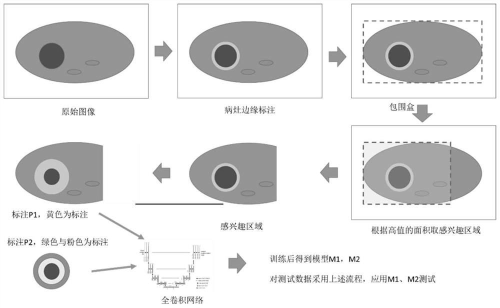
[0029] A method for automatic detection and segmentation of choledochal cyst lesions in abdominal CT, referring to figure 1 , the method includes the following steps:

[0030] Step 1, CT image preprocessing

[0031] 1-1. The doctor marks the edge of the choledochal cyst lesion on the CT source images of the training set to form a closed curve

[0032] 1-2. Perform preliminary processing on the CT source images of the training set and the labels marked by doctors:

[0033] Grayscale mapping: use the sampling window width and window level as the standard (window level: 40, window width: 250) to map the gray value of the data to 0-255.

[0034] Step 2, region of interest extraction

[0035] Since the location of the choledochal cyst is relatively fixed, generally located near the lower part of the liver, but the size of the lesion varies greatly from patient to patient, so we extracted a larger region of interest containing the lesion. Specific steps are as follows:

[0036]...

Embodiment 2

[0056] This embodiment also provides a fully automatic detection and segmentation system for choledochal cyst lesions in abdominal CT images, which uses the method described in Embodiment 1 to detect and segment choledochal cyst lesions in abdominal CT images.

[0057] This embodiment also provides a storage medium on which a computer program is stored, and the program is used to implement the method described in Embodiment 1 when executed.

[0058] This embodiment also provides a computer device, including a memory, a processor, and a computer program stored on the memory and operable on the processor. When the processor executes the computer program, the computer program described in Embodiment 1 is implemented. Methods.


PUM


Abstract

The invention discloses a full-automatic detection and segmentation method and system for common bile duct cyst lesions in abdominal CT. The method comprises the following steps: step 1, conducting CTimage preprocessing; 2, extracting a region of interest; 3, training a network model; 4, performing test segmentation on a CT source image to be segmented; 5, carrying out post-fruiting treatment; and 6, conducting focus edge segmentation. Full-automatic segmentation of common bile duct cyst lesions can be achieved, clinicians can be helped to observe the common bile duct cyst lesions, doctors are helped to formulate treatment strategies, the workload and operation time of the doctors are greatly reduced, and help is provided for follow-up work.

Description

technical field [0001] The invention relates to the technical field of medical image processing, in particular to a method and system for fully automatic detection and segmentation of choledochal cyst lesions in abdominal CT. Background technique [0002] Choledochal cyst is a rare abnormality of the biliary tract whose etiology is unknown, but because it occurs so often in newborns and children it is often thought to be congenital, the most commonly accepted theory being an association with the pancreaticobiliary duct An abnormality of the confluence is associated because it is often observed co-occurring with abnormalities at the common pancreaticobiliary junction. The pancreatic duct and the common bile duct meet outside the ampulla of the hepatopancreatic duct to form a long common channel. The long common channel theory (The long common channel theory) explains that an abnormal junction of the common pancreaticobiliary duct allows the reflux of pancreatic enzymes into t...

Claims


Application Information

Patent Timeline
Patent Type & Authority Applications(China)
IPC IPC(8): G06T7/00G06T7/11
CPCG06T7/0012G06T2207/10081G06T2207/20081G06T2207/20104G06T2207/30096G06T7/11
Inventor 戴亚康耿安康郭万亮耿辰周志勇
Owner SUZHOU INST OF BIOMEDICAL ENG & TECH CHINESE ACADEMY OF SCI
Why Eureka
Try Eureka