Intracranial blood vessel image fusion method and computer readable storage medium

A technology of image fusion and intracranial blood vessels, applied in the field of image processing, can solve the problems of increasing processing difficulty and processing time, different levels of magnetic resonance imaging, unfavorable comprehensive information for doctors, etc., and achieve the effect of accurate diagnosis of intracranial diseases

Inactive Publication Date: 2021-03-16
XIAN CREATION KEJI CO LTD
View PDF0 Cites 4 Cited by
  • Summary
  • Abstract
  • Description
  • Claims
  • Application Information

AI Technical Summary

Problems solved by technology

According to the brightness and darkness of the blood in the magnetic resonance image, the scanning sequence can be divided into bright blood sequence and black blood sequence, but the scanning directions of the black blood sequence and the bright blood sequence are different, resulting in different layers of the final mag

Method used


Image

  • Intracranial blood vessel image fusion method and computer readable storage medium
  • Intracranial blood vessel image fusion method and computer readable storage medium
  • Intracranial blood vessel image fusion method and computer readable storage medium

Examples


Embodiment Construction

[0046] The present invention will be described in further detail below in conjunction with specific examples, but the embodiments of the present invention are not limited thereto.

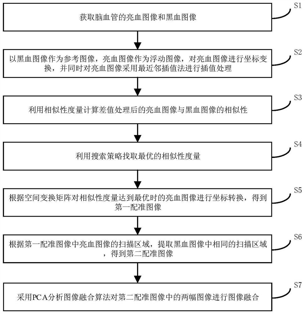
[0047] See figure 1 , figure 1 It is a flowchart of an intracranial blood vessel image fusion method provided by an embodiment of the present invention, as shown in figure 1 As shown, the intracranial blood vessel image fusion method in the embodiment of the present invention includes:

[0048] S1. Acquiring bright blood images and black blood images of intracranial blood vessels.

[0049] At present, methods based on lumen imaging are usually used to evaluate the degree of intracranial vascular lesions and vascular stenosis clinically, such as digital subtraction angiography (Digital Subtraction Angiography, DSA), CT angiography (Computed Tomography Angiography, CTA). ) and high-resolution magnetic resonance angiography (High-Resolution Magnetic Resonance Angiography, HRMRA). The image in the ...


PUM


Abstract

The invention discloses an intracranial blood vessel image fusion method. The method comprises the following steps: acquiring a bright blood image and a black blood image of an intracranial blood vessel; taking the black blood image as a reference image and the bright blood image as a floating image, performing coordinate transformation on the bright blood image, and performing interpolation processing on the bright blood image by adopting a nearest neighbor interpolation method; calculating the similarity between the bright blood image and the black blood image after interpolation processingby using similarity measurement; finding the optimal similarity measurement by using a search strategy, and stopping iteration when the similarity measurement is optimal; according to the space transformation matrix, carrying out coordinate transformation on the bright blood image when similarity measurement is optimal, and obtaining a first registration image; extracting the same scanning area inthe black blood image according to the scanning area of the bright blood image in the first registration image to obtain a second registration image; and adopting a PCA analysis image fusion algorithm to perform image fusion on the two images in the second registration image. According to the scheme, a doctor can be assisted in accurate intracranial disease diagnosis.

Description

technical field [0001] The invention relates to the technical field of image processing, and specifically relates to a fusion method of an intracranial blood vessel image and a computer-readable storage medium. Background technique [0002] With the development of medical imaging technology, a variety of acquisition devices and imaging modes of medical images are emerging, which also brings the need for effective combination of medical images obtained from different medical devices or different acquisition modes of the same medical device. Promote the development of medical image registration and fusion technology. [0003] In particular, the intracranial arteries originate from the carotid and vertebral arteries, which anastomose into the circle of Willis at the base of the brain. The structure is special, the shape is tortuous, and the arterial wall is extremely thin, similar to veins of the same thickness in other parts of the brain; for complex structures Magnetic reson...

Claims


Application Information

Patent Timeline
IPC IPC(8): G06T7/00G06T7/13G06T7/136G06T7/33G06T3/40G06T5/50G06K9/62G16H30/20
CPCG06T7/0012G06T7/13G06T7/136G06T7/337G06T3/4007G06T5/50G16H30/20G06T2207/10088G06T2207/20221G06T2207/30101G06F18/22G06F18/2135
Inventor 石文
Owner XIAN CREATION KEJI CO LTD
Why Eureka
Try Eureka