Systems and methods for magnetic resonance black-blood thrombus imaging in detection of cerebral venous thrombosis

a magnetic resonance and black blood thrombosis technology, applied in the field of magnetic resonance imaging methods, can solve the problems of difficult to exclude cvt with existing noninvasive imaging modalities, death or permanent disability, and the frequency of unrecognized cvt, and achieve the effect of accelerating imaging speed

Inactive Publication Date: 2017-08-10
CEDARS SINAI MEDICAL CENT
View PDF1 Cites 2 Cited by
  • Summary
  • Abstract
  • Description
  • Claims
  • Application Information

AI Technical Summary

Benefits of technology

[0006]In various embodiments, the invention teaches a method for performing magnetic resonance imaging (MM) on a subject, including (1) acquiring magnetic resonance data from a volume of interest (VOI) that includes a blood vessel of a subject's head and/or neck, by using a 3D variable-flip-angle turbo spin-echo (TSE) acquisition, and (2) generating one or more images based on said data. In some embodiments, the blood vessel includes a thrombus. In certain embodiments, the thrombus is depicted as hyperintense compared with surrounding tissues. In some embodiments, the subject has cerebral venous thrombosis (CVT). In some embodiments, the method further includes using T1-weighting. In some embodiments, the MRI machine used to perform the magnetic resonance imaging is a 1.5 T scanner or a 3.0 T scanner. In various embodiments, the VOI includes one or more of the following anatomical structures or regions within the subject: superior sagittal sinus, right transverse sinus, right sigmoid sinus, left transverse sinus, left sigmoid sinus, straight sinus, confluence of sinuses, veins of galen, internal cerebral veins, veins of Labbé, right cortical veins, and left cortical veins. In some embodiments, the subject is a human.
[0007]In various embodiments, the invention teaches a magnetic resonance imaging (MRI) system that includes (1) a magnet operable to provide a magnetic field; (2) a transmitter operable to transmit to a region within the magnetic field; (3) a receiver operable to receive a magnetic resonance signal from the region; and (4) a processor operable to control the transmitter and the receiver; wherein the processor is configured to direct the transmitter and receiver to execute a sequence that includes (a) acquiring magnetic resonance data from a blood vessel within a volume of interest (VOI) that includes all or a portion of a subject's head and/or neck, according to the me

Problems solved by technology

However, CVT is frequently unrecognized and the average delay from the onset of symptoms to the diagnosis is about one week, because the positive findings of intraluminal thrombus are not always evident.
However, given the variation in venous anatomy, it is sometimes difficult to exclude CVT with existing noninvasive imaging modalities.
The diagnosis dilemma may delay treatment and result in death or permanent disability.
There are still m

Method used


Image

  • Systems and methods for magnetic resonance black-blood thrombus imaging in detection of cerebral venous thrombosis
  • Systems and methods for magnetic resonance black-blood thrombus imaging in detection of cerebral venous thrombosis
  • Systems and methods for magnetic resonance black-blood thrombus imaging in detection of cerebral venous thrombosis

Examples


example 1

Subjects and Methods

Patients

[0030]Between February 2014 and May 2015, consecutive patients with signs and symptoms suspected of CVT within the past 30 days were prospectively recruited. Exclusion criteria included general contraindications to MR examination and patients with incomplete conventional imaging examinations (CT, MR, and MRV). Informed consent was obtained from all participants, and all protocols were approved by the Institutional Review Board.

Conventional Imaging Evaluation

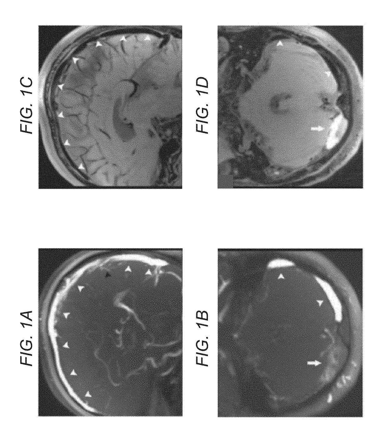
[0031]Thrombi were defined as intraluminal filling defects detected by conventional imaging techniques. Two readers (J.D. and X.J.) performed a consensus reading of all conventional imaging studies for each patient, including CT, MR, and MRV, with full clinical and outcome information on the patient to obtain a reference standard. The following 14 venous segments were included in evaluations: superior sagittal sinus, inferior sagittal sinus, right transverse sinus, right sigmoid sinus, left transverse ...


PUM


Abstract

In various embodiments, the present invention teaches systems and methods for using T1-weighted black-blood MR imaging, with which a CVT can be well isolated from the surrounding tissues due to the signal suppression of flowing blood. In some embodiments, the invention teaches using black-blood imaging (3D variable-flip-angle turbo spin-echo acquisition) to directly visualize thrombi. In certain embodiments, the invention teaches using T1 weighted image contrast and isotropic sub-millimeter spatial resolution for accurate detection and staging of thrombi. In various embodiments, the invention allows for the detection of chronic thrombosis recanalization.

Description

FIELD OF THE INVENTION[0001]The present invention generally relates to imaging methods, and especially magnetic resonance imaging methods.BACKGROUND[0002]The following description includes information that may be useful in understanding the present invention. It is not an admission that any of the information provided herein is prior art or relevant to the presently claimed invention.[0003]Cerebral venous thrombosis (CVT), including thrombosis of cerebral veins and major dural sinuses, is a form of stroke that usually affects young individuals. During the past decade, improved diagnosis and treatments have improved the outcome of CVT. However, CVT is frequently unrecognized and the average delay from the onset of symptoms to the diagnosis is about one week, because the positive findings of intraluminal thrombus are not always evident. Diagnosis of CVT typically relies on a combination of different imaging modalities, such as computed tomography (CT) venography, magnetic resonance (M...

Claims


Application Information

Patent Timeline
IPC IPC(8): A61B5/00A61B5/055
CPCA61B5/0042A61B2576/026A61B5/4064A61B5/055G01R33/5607G01R33/4822G01R33/5617A61B5/02007G01R33/5602G16H30/40
Inventor YANG, QIFAN, ZHAOYANGLI, DEBIAOJI, XUNMING
Owner CEDARS SINAI MEDICAL CENT
Why Eureka
Try Eureka