Method and device for detecting nodules in thyroid ultrasound image based on deep learning

A technology for thyroid nodules and ultrasound images, which is applied in image enhancement, image analysis, image data processing, etc., can solve the problems of different sizes of nodule areas in images, waste of computing resources, and increase the amount of computation, so as to improve experimental testing. Indicators, improving test indicators, and enhancing the effect of generalization performance

Pending Publication Date: 2021-04-06
THE FIRST MEDICAL CENT CHINESE PLA GENERAL HOSPITAL +2
View PDF0 Cites 4 Cited by
  • Summary
  • Abstract
  • Description
  • Claims
  • Application Information

AI Technical Summary

Problems solved by technology

[0003] However, the prior art solution has the following two disadvantages: (1) It is necessary to set three anchor boxes with sizes of 64, 128 and 256 and three ratios of 1:2, 1:1 and 2:1 before the experiment, however , due to the actual situation, the size of the input thyroid ultrasound image is not fixed, and the size of the nodule area in the image is different. The hyperparameters such as the size, aspect ratio, and number of the anchor box will have a great impact on the experimental results. In Faced with a huge test in the process of actual auxiliary diagnosis
(2) In order to bring a higher recall rate to the experimental results, a large num

Method used


Image

  • Method and device for detecting nodules in thyroid ultrasound image based on deep learning
  • Method and device for detecting nodules in thyroid ultrasound image based on deep learning
  • Method and device for detecting nodules in thyroid ultrasound image based on deep learning

Examples


Embodiment Construction

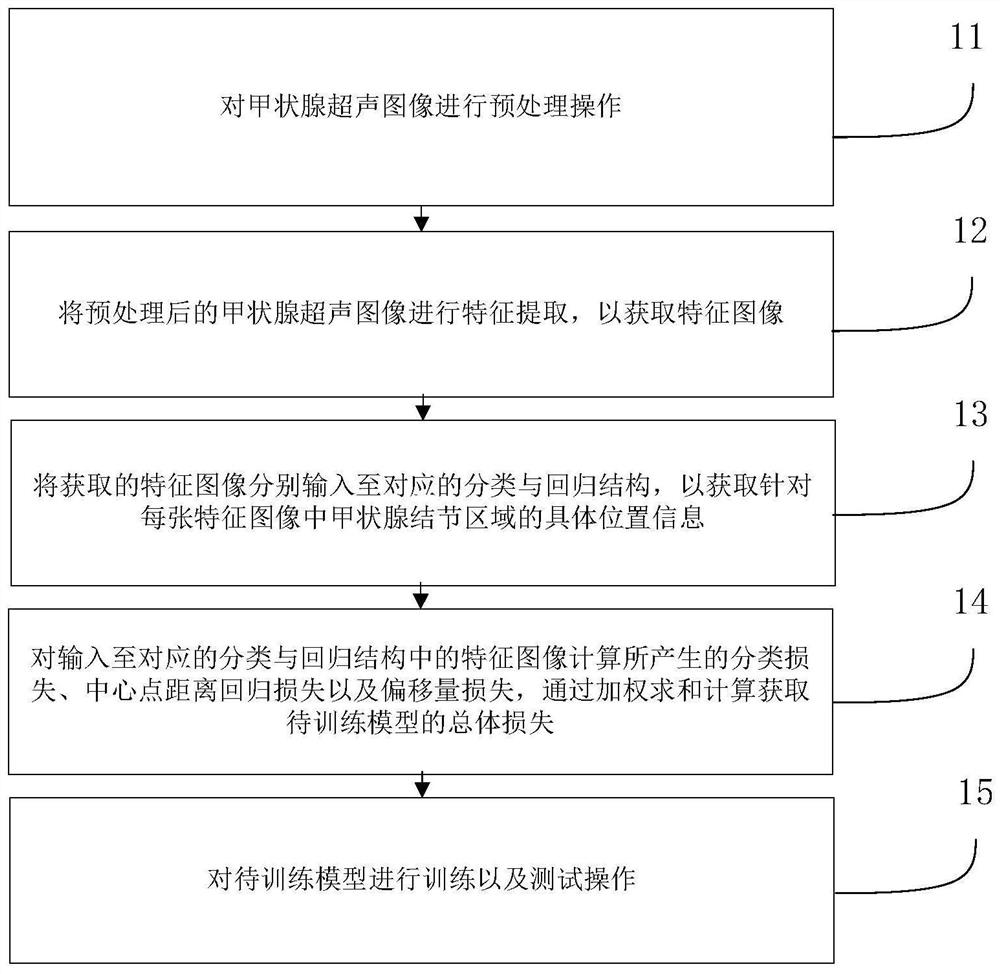
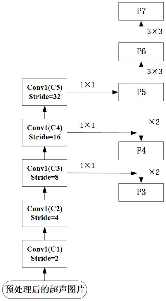
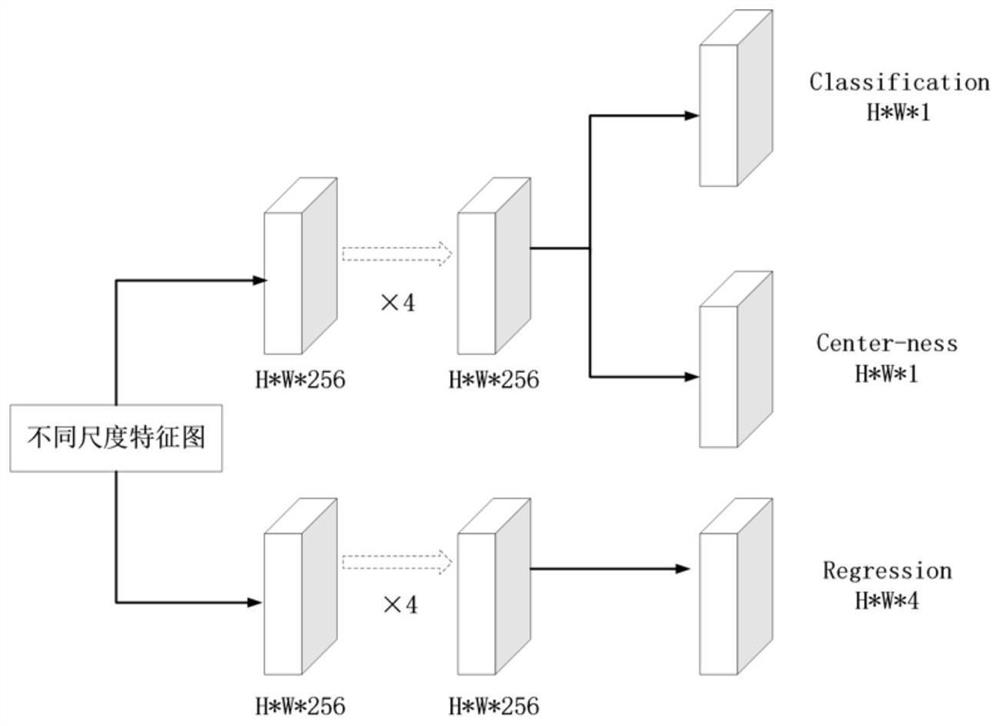
[0035] The present application will be further described in detail below in conjunction with the accompanying drawings and embodiments.

[0036]In the following introduction, the terms "first" and "second" are only used for the purpose of description, and should not be understood as indicating or implying relative importance. The following introduction provides multiple embodiments of the present disclosure, and different embodiments can be replaced or combined and combined, so the application can also be considered to include all possible combinations of the same and / or different embodiments described. Thus, if one embodiment contains features A, B, C, and another embodiment contains features B, D, then the application should also be considered to include all other possible combinations containing one or more of A, B, C, D Although this embodiment may not be clearly written in the following content.

[0037] In order to make the purpose, technical solution and advantages of ...


PUM


Abstract

The invention provides a method for detecting nodules in a thyroid ultrasound image based on deep learning. The method comprises the steps of preprocessing the thyroid ultrasound image; extracting features of the preprocessed thyroid ultrasound image to obtain a feature image; respectively inputting the obtained feature images into corresponding classification and regression structures, and obtaining specific position information of a thyroid nodule region in each feature image; for the classification loss, the central point distance regression loss and the offset loss generated by calculation of the feature images input into the corresponding classification and regression structures, obtaining the total loss of the to-be-trained model through weighted summation calculation; and training and testing the to-be-trained model. According to the method, an anchor box does not need to be arranged, the nodule region in the thyroid ultrasound image is efficiently detected, calculation and resource waste related to the anchor box are avoided, the training speed is increased, and the generalization performance of an experimental result is enhanced. The invention further provides a device for detecting the nodules in the thyroid ultrasound image based on deep learning.

Description

technical field [0001] The present disclosure relates to the technical field of computer biology, in particular, to a method and device for detecting nodules in thyroid ultrasound images based on deep learning. Background technique [0002] At present, there are many technologies that use deep learning methods for auxiliary diagnosis on medical images. For the auxiliary diagnosis of thyroid nodules in thyroid ultrasound images, the commonly used deep learning methods can be divided into two categories: one is the dual network based on the region candidate network. stage detector, and the other is a single-stage detector that is not based on a region candidate network. In general, the two methods require the following steps: firstly, obtain the feature map of the ultrasound image after the features are extracted by the convolutional neural network; secondly, set the anchor point (anchor) and a certain number and size of the anchor box (anchor box) on the feature map ; Finall...

Claims


Application Information

Patent Timeline
IPC IPC(8): G06T7/00
CPCG06T7/0012G06T2207/10132G06T2207/30004G06T2207/20081G06T2207/20084
Inventor 罗渝昆谢芳林科汝陈东浩张艳田晓琦张颖王筱斐叶丹任改霞李发根欧阳勇春
Owner THE FIRST MEDICAL CENT CHINESE PLA GENERAL HOSPITAL
Why Eureka
Try Eureka