Intestinal disease segmentation method of salient edge feature extraction module guided network

An edge feature, guiding network technology, applied in neural learning methods, biological neural network models, image analysis, etc., can solve problems such as inaccurate target positioning and image segmentation errors, and achieve the effect of improving segmentation performance and large differences in shape changes.

Pending Publication Date: 2021-04-13
ZHEJIANG UNIV OF TECH
View PDF0 Cites 8 Cited by
  • Summary
  • Abstract
  • Description
  • Claims
  • Application Information

AI Technical Summary

Problems solved by technology

Improved the problem that the PraNet network mis-segmented images containing light spots due t

Method used


Image

  • Intestinal disease segmentation method of salient edge feature extraction module guided network
  • Intestinal disease segmentation method of salient edge feature extraction module guided network
  • Intestinal disease segmentation method of salient edge feature extraction module guided network

Examples


Embodiment Construction

[0026] In order to illustrate the purpose, technical solutions and advantages of the present invention, the present invention will be further described in detail below in conjunction with specific embodiments and accompanying drawings.

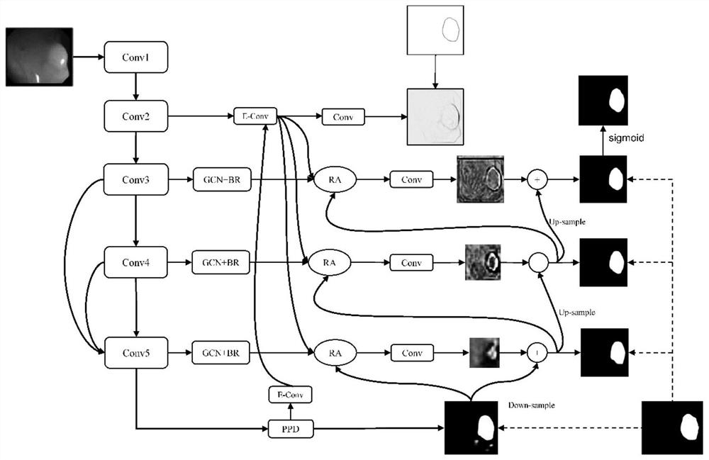
[0027] refer to Figure 1 to Figure 6 , a salient edge feature extraction module guided network intestinal disease segmentation method, including the following steps:

[0028] Step 1: Input data set X={x 1 ,x 2 ,...,x n}, where X represents the input samples in the data set,

[0029] x n ∈ R 352×352 , n represents the number of samples, the backbone network Res2net extracts features, and obtains five output features of Conv1, Conv2, Conv3, Conv4, and Conv5;

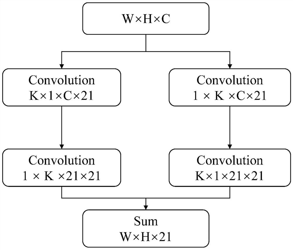
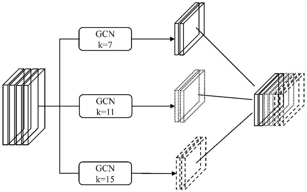
[0030] Step 2: Due to the size and shape of polyps in the colorectal polyp data set vary greatly, GCN modules with different kernel sizes are used to extract multi-scale context information, and the boundary refinement module (BR module) is used to further improve the accuracy of ob...


PUM


Abstract

An intestinal disease segmentation method of a significant edge feature extraction module guided network comprises the steps of: firstly, inputting a data set, extracting features through a backbone network Res2net, then extracting multi-scale context information through GCN modules with different kernel sizes, improving the division performance of object boundaries through a boundary refinement module, and further extracting the features; generating edge information by using the second-layer side edge output features, suppressing noise in the edge information by using the three fused high-layer semantic information, and performing significant edge supervision on the generated significant edge features by using a mask image generation edge image to form a final significant edge feature extraction module; and finally, jointly inputting the edge information generated in the significant edge feature extraction module and the side output information of each layer into the RAS module, and training the model to obtain training parameters, wherein the training parameters are used for testing to obtain a final result. According to the invention, the problems of inaccurate polyp segmentation and positioning and fuzzy boundary in gastrointestinal tracts are improved.

Description

technical field [0001] The invention relates to the technical field of image processing of artificial intelligence, in particular to a method for segmenting intestinal diseases with a salient edge feature extraction module-guided network. Background technique [0002] Colorectal cancer is the third most common cause of cancer-related death worldwide and usually results from the abnormal growth of polyps in the colon. Colonoscopy is the main method for screening and preventing polyp canceration. However, colonoscopy relies on highly skilled endoscopists and a high degree of eye-hand coordination. Studies have shown that the polyp missed rate in patients undergoing colonoscopy reaches 22%. %-28%. Segmenting polyps from normal mucosa can help endoscopists improve mis-segmentation and subjectivity. In order to accurately segment polyps, many different methods have been proposed. Existing polyp segmentation research work can be roughly summarized into three main methods, the f...

Claims


Application Information

Patent Timeline
IPC IPC(8): G06T7/12G06T7/181G06T5/00G06T5/50G06K9/62G06N3/04G06N3/08
CPCG06T7/12G06T7/181G06T5/002G06T5/50G06N3/08G06T2207/10068G06T2207/30032G06T2207/20221G06N3/045G06F18/253
Inventor 李胜夏瑞瑞何熊熊程珊郝明杰王栋超
Owner ZHEJIANG UNIV OF TECH
Why Eureka
Try Eureka