Imaging device through slit scanning

A technology of imaging equipment and slit scanning, which is used in the control of radiological diagnostic equipment, instruments used for radiological diagnosis, medical science, etc. It can solve the problems of splicing error, point light source imaging amplification, insufficient quantitative analysis, etc., and avoid the amplification effect. Effect

Pending Publication Date: 2021-05-07
SHANGHAI TAOIMAGE MEDICAL TECH CO LTD
View PDF7 Cites 1 Cited by
  • Summary
  • Abstract
  • Description
  • Claims
  • Application Information

AI Technical Summary

Problems solved by technology

[0002] At present, medical imaging technology has been widely used clinically. Conventional X-ray plain film is the most important inspection method. At present, digital X-ray photography systems are equipped in major hospitals, which play an indispensable role in the qualitative diagnosis of diseases. , but the quantitative analysis is somewhat insufficient, due to the point light source imaging has the effect of magnifying the object, so obtaining the image of the true proportion of the patient has always been a clinical pursuit
In the prior art, the receiver is a flat panel, and a rectangular image is captured, which is stitched based on several rectangular images. Due to the difference in magnification, there are errors in the stitching

Method used


Image

  • Imaging device through slit scanning
  • Imaging device through slit scanning
  • Imaging device through slit scanning

Examples


Embodiment

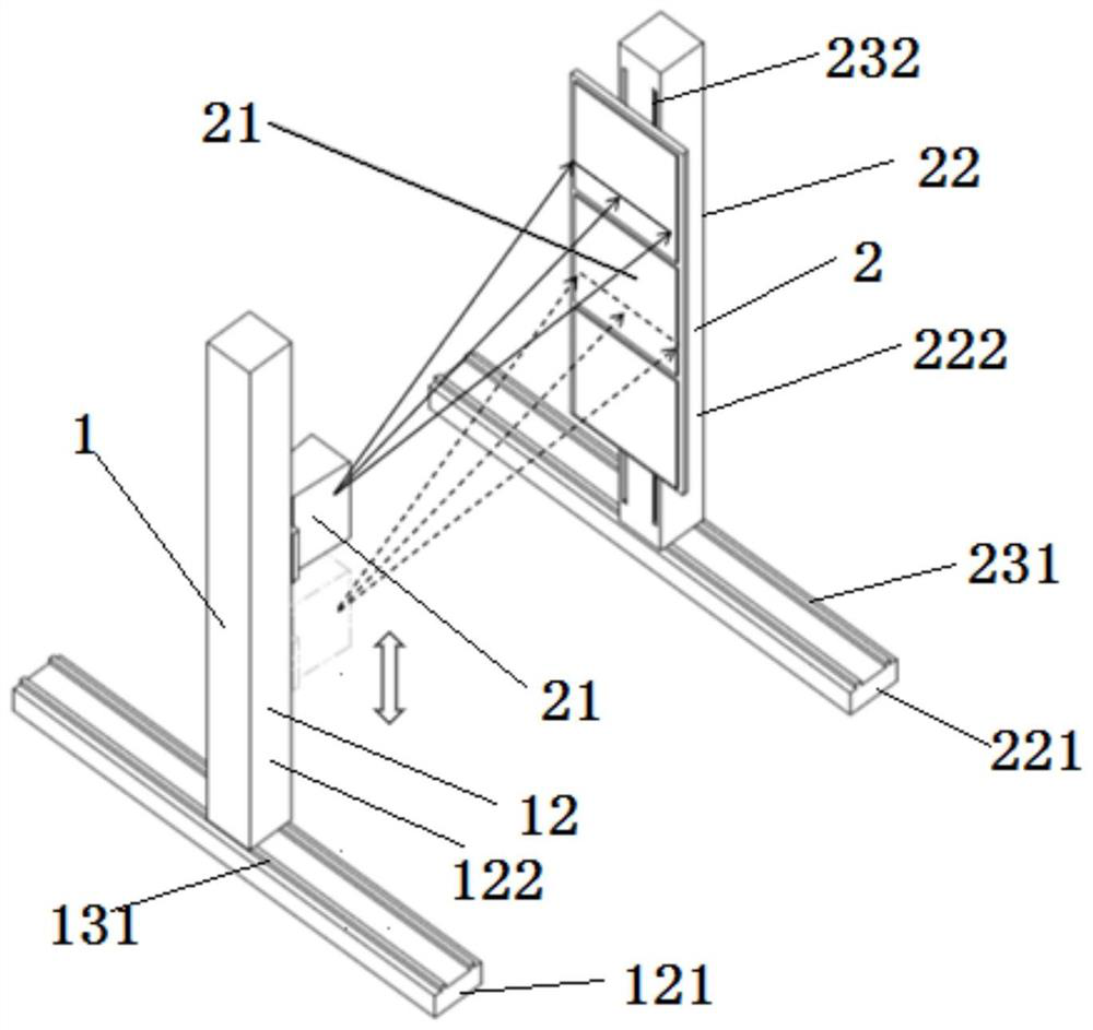
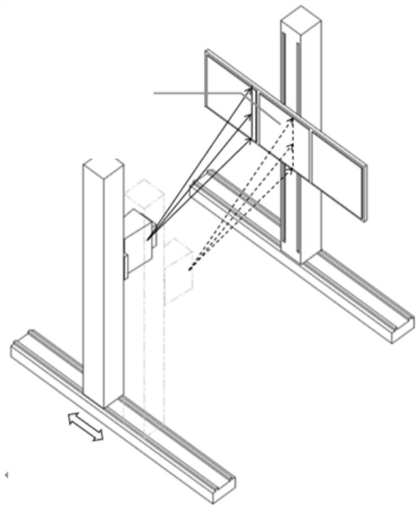
[0056] Such as figure 1 with figure 2 As shown, this embodiment provides an imaging device that scans through a slit, including: a transmitting end 1 and a receiving end 2, wherein the transmitting end 1 is arranged opposite to the receiving end 2;

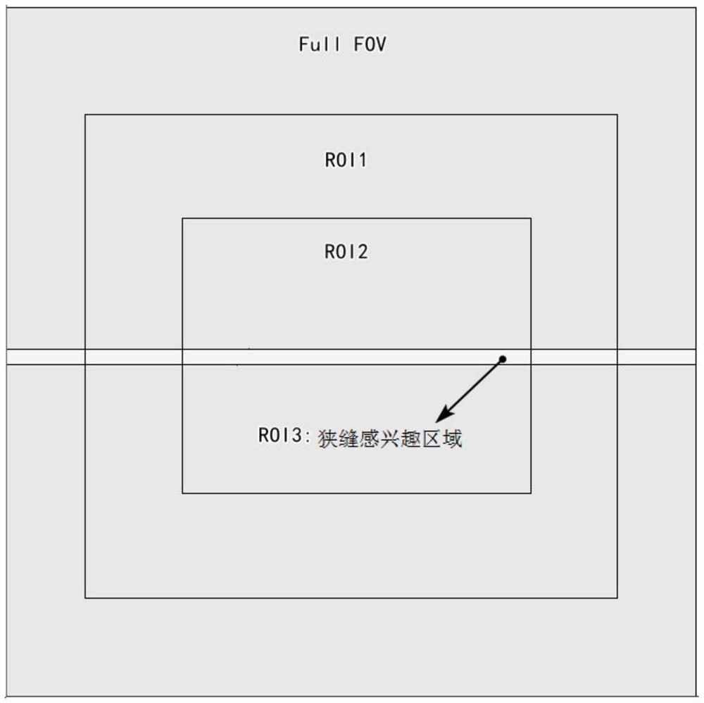
[0057] The emitting end 1 is used to collimate the X-rays into slit X-rays, continuously send the slit X-rays moving in any way including up and down, left and right to the object to be imaged, and linearly move into slit X-rays. The way of making the object to be imaged is completely photographed by the slit X-ray; Activate the slit ROI corresponding to the slit X-ray emission direction on the receiving end 2, obtain the attenuated slit X-ray through the slit ROI, and convert it into The slit images with the same X-ray shooting range are stored, and after the object to be imaged is completely shot, all the slit images are spliced ​​to form a complete imaging picture of the object to be imaged.

[0058] Specifically, in tradit...


PUM


Abstract

The invention relates to the technical field of digital X-ray imaging methods, and discloses an imaging device through slit scanning. The imaging device comprises a transmitting end and a receiving end, wherein the transmitting end is used for collimating X-rays into slit X-rays, continuously transmitting the slit X-rays to an object to be imaged, and completely shooting the object to be imaged by the slit X-rays in a linear motion surface forming mode; and the receiving end is used for automatically activating a slit region of interest corresponding to the transmitting direction of the slit X-rays on the receiving end after the slit X-rays transmitted each time pass through the object to be imaged, acquiring attenuated slit X-rays, converting the slit X-rays into slit images with the same shooting range as the slit X-rays for storage, and splicing the slit images after complete shooting. The X-rays are collimated in a slit scanning mode, so that the amplification effect of point light source imaging is avoided; the imaging device can be used for acquiring images of a patient in a real proportion; and the radiation dose of the patient is far lower than the dose of spiral scanning, and is at the level of common radiation.

Description

technical field [0001] The invention relates to the technical field of digital X-ray imaging methods, in particular to an imaging device that scans through a slit. Background technique [0002] At present, medical imaging technology has been widely used clinically. Conventional X-ray plain film is the most important inspection method. At present, digital X-ray photography systems are equipped in major hospitals, which play an indispensable role in the qualitative diagnosis of diseases. , but it seems to be somewhat insufficient for quantitative analysis, due to the fact that point light source imaging has the effect of magnifying objects, so obtaining images of the true proportion of patients has always been a clinical pursuit. In the prior art, the receiver is a flat panel, and a rectangular image is captured, which is stitched based on several rectangular images. Due to the difference in magnification, there are errors in the stitching. [0003] With the development of te...

Claims


Application Information

Patent Timeline
Patent Type & Authority Applications(China)
IPC IPC(8): A61B6/00A61B6/06
CPCA61B6/44A61B6/06A61B6/40A61B6/42A61B6/4476A61B6/4429A61B6/54
Inventor 蔡宗远白鹏鹏齐彦君周卫东
Owner SHANGHAI TAOIMAGE MEDICAL TECH CO LTD
Why Eureka
Try Eureka