Method and device for recognizing echocardiogram of congenital heart disease

A technology of congenital heart disease and echocardiography, applied in neural learning methods, character and pattern recognition, image enhancement, etc., can solve inaccurate ultrasound image positioning, high misdiagnosis rate, and congenital heart disease identification depends on doctor identification Ability and other issues, to achieve the effect of low cost, easy method and wide applicability

Pending Publication Date: 2021-05-07
BEIJING CHILDRENS HOSPITAL AFFILIATED TO CAPITAL MEDICAL UNIV +1
View PDF0 Cites 3 Cited by
  • Summary
  • Abstract
  • Description
  • Claims
  • Application Information

AI Technical Summary

Benefits of technology

This patented technology allows for easier identification of cardiovascular conditions through analysis on X rays or ultrasound images obtained from patients' blood vessels during surgery procedures. It also helps diagnose certain types of abnormalities such as valve regurgitation (VTR) caused due to genetic defects that affect how much fluid flows between two chambers inside the body.

Problems solved by technology

This patented technical problem addressed by this patents relates to improving diagnosis for CHDs due to their unique characteristics that make it difficult or impossible to accurately locate them during an examination process. Ultrasonic imagery has been shown effective but only at specific locations within certain areas where there can be significant differences between patients' hearts compared to those from other parts of the body. Therefore, these techniques require trained professionals who work alongside experts to recognize even small variations caused by changes made over time through medical procedures like cardiac catheterization.

Method used


Image

  • Method and device for recognizing echocardiogram of congenital heart disease
  • Method and device for recognizing echocardiogram of congenital heart disease
  • Method and device for recognizing echocardiogram of congenital heart disease

Examples


specific Embodiment 1

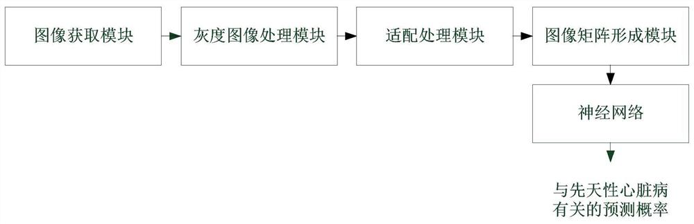
[0053] 1. Image acquisition

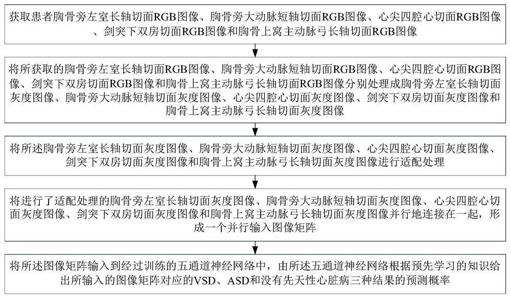
[0054] The images used in this application were collected by Philips Xinyue IE33 cardiac color Doppler ultrasound, Philips IE Elite cardiovascular four-dimensional color Doppler ultrasound and Philips Epiq7C ultra-high-end advanced cardiac color Doppler ultrasound equipment. The collected images are parasternal left ventricle long-axis section view, parasternal aorta short-axis Section view, apical four-chamber view, subxiphoid-process biatrial view, and suprasternal fossa aortic arch long-axis view, wherein the images are RGB images.

[0055] 2. Image processing

[0056] The collected parasternal left ventricle long-axis view, parasternal aorta short-axis view, apical four-chamber view, subxiphoid-process double-chamber view, and suprasternal fossa aortic arch long-axis view were processed in grayscale and rectangularly cropped Keep the target area. Since the acquired echocardiogram is fan-shaped, some target areas cannot be cropped by rectangl...

specific Embodiment 2

[0063] 1. Acquisition of echocardiographic data

[0064] 330 cases of healthy controls, 145 cases of VSD and 91 cases of ASD were collected from Beijing Children's Hospital. Put the subject in a supine position, expose the chest, use the instrument on the subject's heart according to the instructions, and collect the parasternal left ventricular long-axis view, parasternal aorta short-axis view, and apical four-chamber heart section of each subject. Standard two-dimensional views of the plane, the subxiphoid-process bicameral view, and the suprasternal fossa long-axis view of the aortic arch.

[0065] The diagnosis of all subjects was confirmed by at least two senior ultrasound doctors or the final diagnosis during the operation, and the operation was approved by the Ethics Committee of Beijing Children's Hospital (approval number: 2019-k-342).

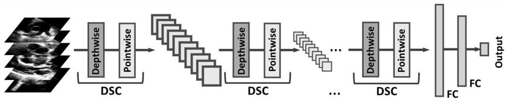
[0066] 2. Image preprocessing

[0067] The three-channel RGB image of the section image obtained in step 1 is processed into a sin...


PUM


Abstract

The invention discloses an echocardiogram recognition method for congenital heart disease. The method comprises the following steps: collecting five RGB images of a left greenhouse long-axis section RGB image beside the sternum, a main artery short-axis section RGB image beside the sternum, a heart apex four-cavity heart tangent surface RGB image, a xiphoid process lower double-chamber section RGB image and a sternum superior fossa aortic arch long-axis section RGB image of a patient; the five obtained RGB images are respectively processed into five gray level images; carrying out adaptive processing on the five gray level images; connecting the five grayscale images subjected to adaptive processing together in parallel to form a parallel input image matrix; and inputting the image matrix into a trained five-channel neural network, and giving prediction probabilities of three results of VSD, ASD and no congenital heart disease corresponding to the input image matrix by the five-channel neural network according to pre-learned knowledge.

Description


Claims


Application Information

Patent Timeline
Owner BEIJING CHILDRENS HOSPITAL AFFILIATED TO CAPITAL MEDICAL UNIV
Why Eureka
Try Eureka