Eureka AIR delivers breakthrough ideas for toughest innovation challenges, trusted by R&D personnel around the world.

Optical coherence tomography angiography method and device

An optical coherence tomography and angiography technology, which is used in medical science, blood flow measurement, and diagnosis using tomography. Small, improve work efficiency, and reduce algorithm complexity

Pending Publication Date: 2020-07-24
TOWARDPI (BEIJING) MEDICAL TECH LTD
View PDF6 Cites 3 Cited by
  • Summary
  • Abstract
  • Description
  • Claims
  • Application Information

AI Technical Summary

Problems solved by technology

[0005] The present invention provides an optical coherence tomography angiography method and device to solve the problem of weak blood flow signal and high noise in angiography, and it is impossible to obtain relatively clear blood vessel images

Method used

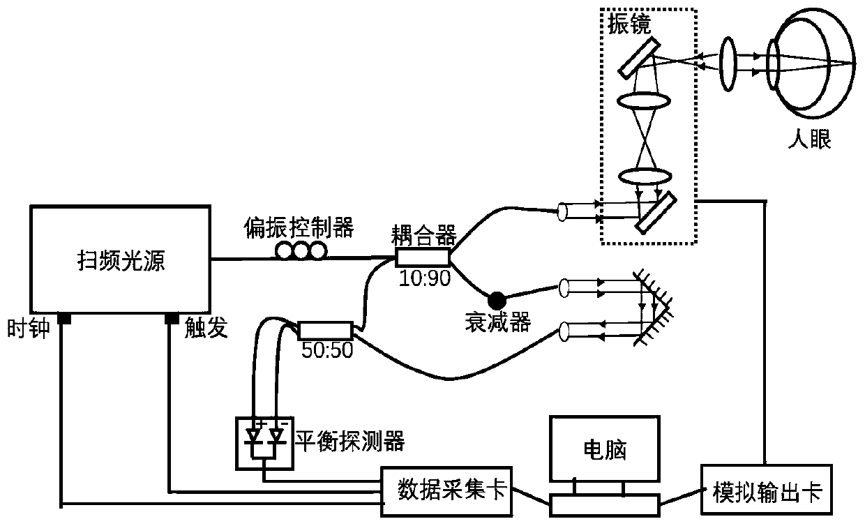
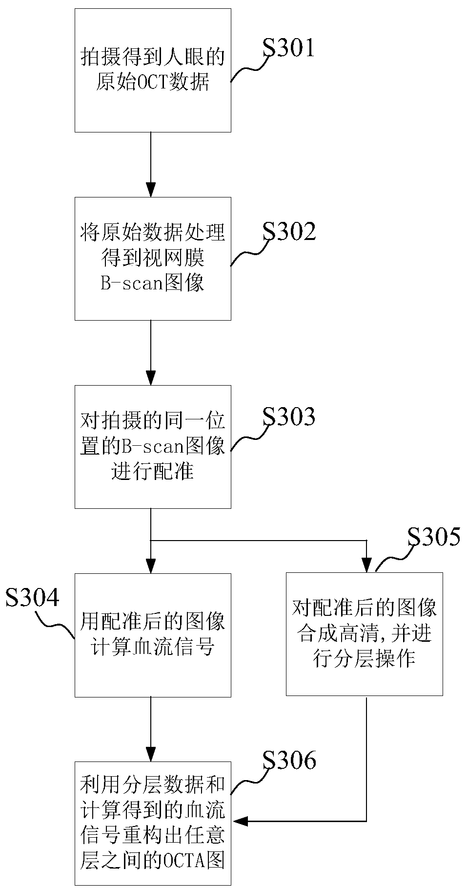

Image

  • Optical coherence tomography angiography method and device
  • Optical coherence tomography angiography method and device
  • Optical coherence tomography angiography method and device

Examples


Embodiment Construction

[0053] Reference will now be made in detail to the exemplary embodiments, examples of which are illustrated in the accompanying drawings. When the following description refers to the accompanying drawings, the same numerals in different drawings refer to the same or similar elements unless otherwise indicated. The implementations described in the following exemplary examples do not represent all implementations consistent with the present invention. Rather, they are merely examples of apparatuses and methods consistent with aspects of the invention as recited in the appended claims.

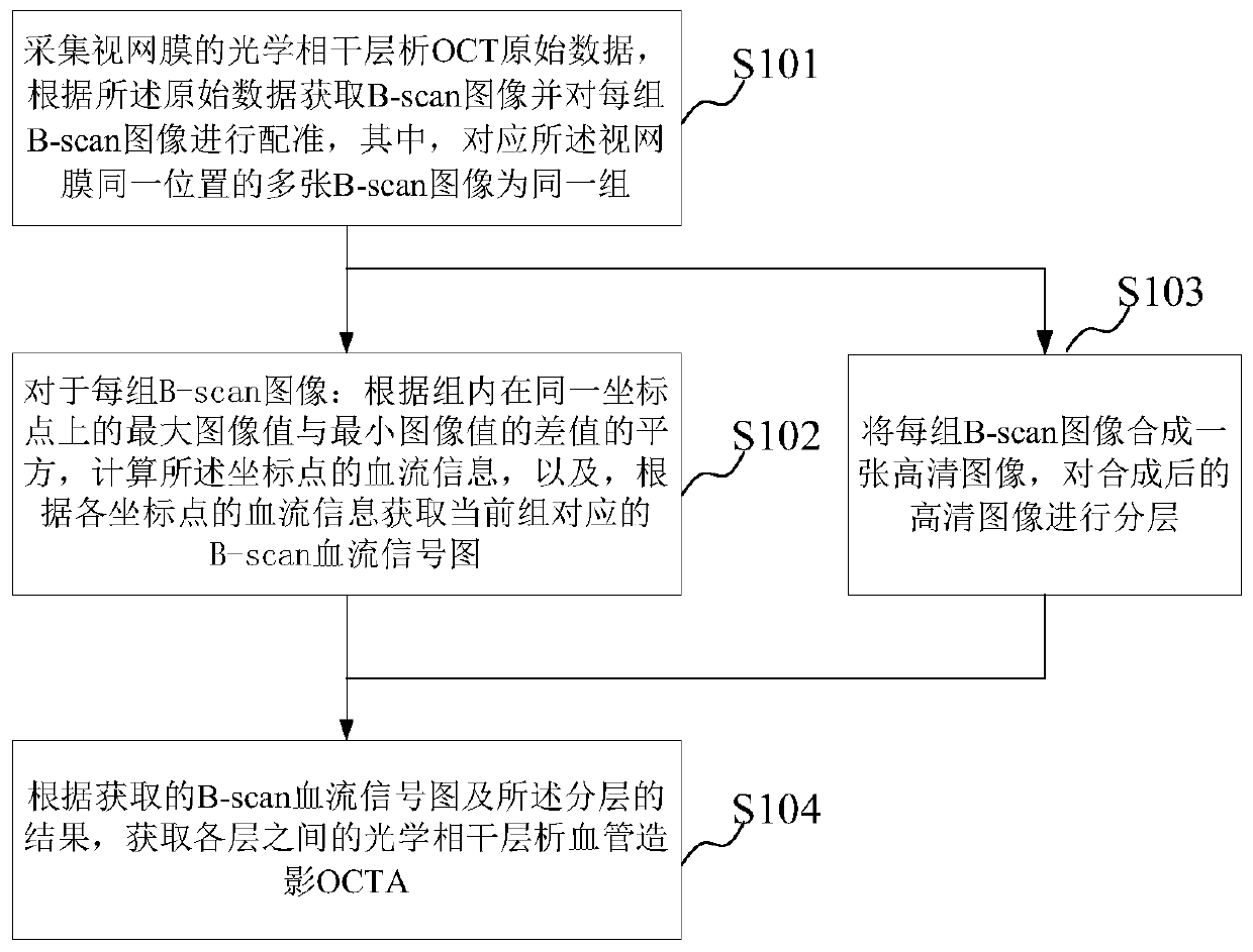
[0054] figure 1 It is a flowchart of an optical coherence tomography angiography method according to an exemplary embodiment of the present invention. The method can be used in a system consisting of OCT equipment, computers, and the like.

[0055] see figure 1 As shown, the method may include:

[0056] Step S101, collecting the original optical coherence tomography OCT data of the retina, o...


PUM


Abstract

The invention provides an optical coherence tomography angiography method and device. The method comprises the steps: collecting OCT original data of a retina, obtaining B-scan images, and carrying out registration on each group of images; calculating blood flow information of a coordinate point according to the square of the difference value of the maximum image value and the minimum image valueon the same coordinate point in each group, and further obtaining a blood flow signal graph corresponding to the current group; synthesizing each group of images into a high-definition image and layering the high-definition images; and , obtaining the OCTA among the layers according to the blood flow signal graph and the layering result. Compared with the prior art, the influence on the noise partis small, the blood flow signal is enhanced, and the discrimination between the blood flow signal and the noise is increased, so that the quality of the obtained angiography image is higher, capillary details are richer, and therefore a doctor can diagnose fundus diseases of a patient easily. Meanwhile, the algorithm complexity is low, the calculation amount is small, the burden of a computer isreduced, the calculation speed of the blood flow signals is increased, and therefore the working efficiency is improved.

Description

technical field [0001] The invention relates to the field of optical coherence tomography, in particular to an optical coherence tomography angiography method and device. Background technique [0002] OCT (Optical Coherence Tomography, optical coherence tomography, or optical coherence tomography) is a high-sensitivity, high-resolution, high-speed, non-invasive imaging method, which has been widely used in the diagnosis of fundus diseases. The detection and treatment of eye diseases is of great significance. As a tomographic imaging method, OCT uses the coherence of light to scan and image the fundus. Each scan is called an A-scan, and the combination of adjacent and continuous scans is called a B-scan. B-scan That is, the commonly seen OCT cross-sectional view is the most important imaging method of OCT in medical diagnosis. [0003] In the field of OCT, OCTA (OCT Angiography, optical coherence tomography angiography) is a new technology developed in recent years. Retinal...

Claims


Application Information

Patent Timeline
IPC IPC(8): A61B5/00A61B5/026
CPCA61B5/0066A61B5/0073A61B5/0261
Inventor 汤剑宇汪霄
Owner TOWARDPI (BEIJING) MEDICAL TECH LTD
Why Eureka
Eureka Blog
Learn More