Multi-section medical image compression method based on three-dimensional discrete wavelet transform

A discrete wavelet transform and medical image technology, applied in image communication, electrical components, digital video signal modification, etc., can solve the problem that it is difficult to compress multiple multi-section medical images at the same time, so as to solve the problem of inter-frame redundancy, eliminate The effect of image square distortion and efficient data transmission

Active Publication Date: 2021-06-18
XIANGTAN UNIV
View PDF15 Cites 0 Cited by
  • Summary
  • Abstract
  • Description
  • Claims
  • Application Information

AI Technical Summary

Problems solved by technology

However, mainstream compression algorithms can only compress a single image, which is difficult to meet the requirements of simultaneously compressing multiple multi-section medical images

Method used


Image

  • Multi-section medical image compression method based on three-dimensional discrete wavelet transform
  • Multi-section medical image compression method based on three-dimensional discrete wavelet transform
  • Multi-section medical image compression method based on three-dimensional discrete wavelet transform

Examples


Embodiment Construction

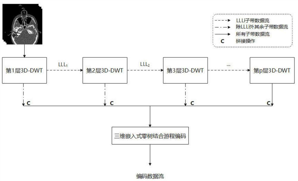
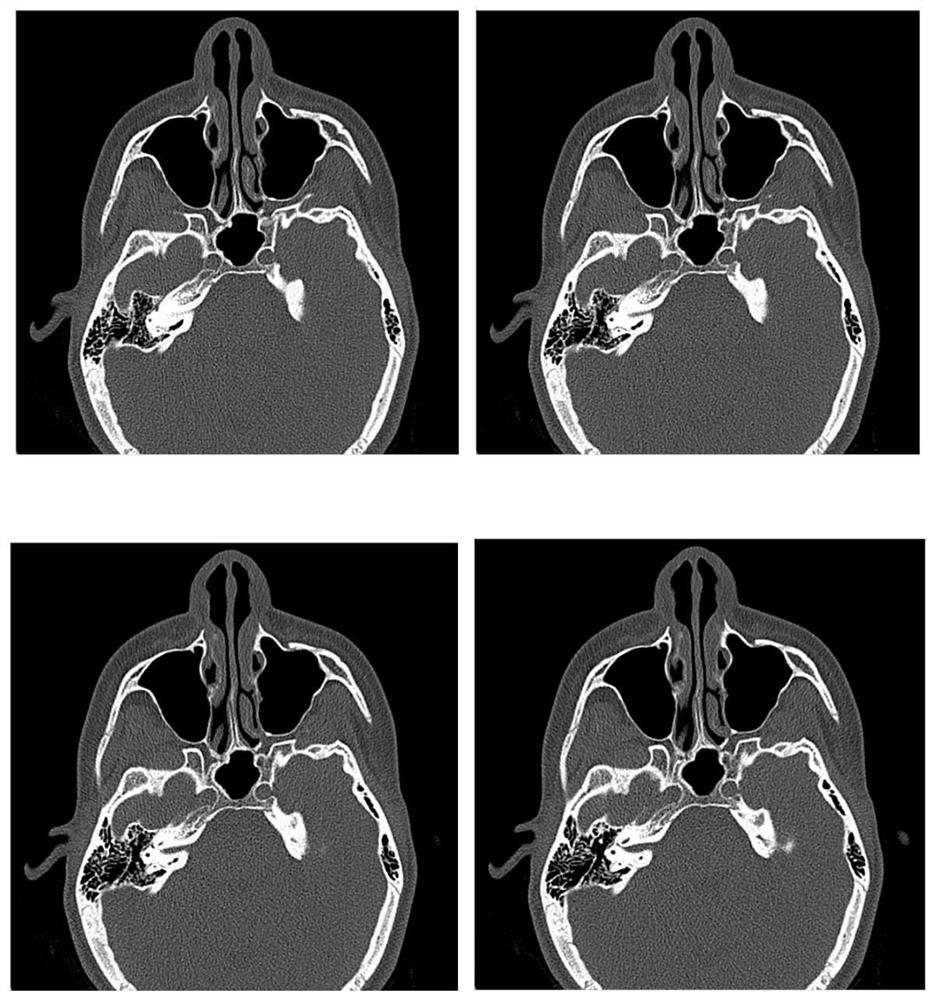
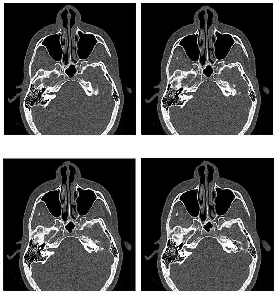
[0020] Implement the present invention, take 8 pieces of human brain CT scanning pictures as picture sources, and MATLAB is the realization environment as an example to illustrate. 8 CT images of the brain such as figure 2 As shown, the CT image is composed of a series of grayscale pixels arranged according to a certain size, and the saving format is bmp. It will be stored in the form of RBG directly read from the file. Before starting to compress, it should be converted into a grayscale image. The subsequent CT image data matrix is ​​512*512, stored in uint8 type.

[0021] combine figure 1 , the concrete implementation steps of the present invention are as follows:

[0022] S100. Read 8 multi-section medical image sources, convert all images into grayscale images, and set the size to 512*512, store the transformed images in the same matrix, and obtain a size of 512*512*8 Three-dimensional image data matrix Image, perform three-layer three-dimensional discrete wavelet tran...


PUM


Abstract

The invention provides a multi-section medical image compression method based on three-dimensional discrete wavelet transform. The method comprises the following steps: firstly, carrying out multi-layer three-dimensional discrete wavelet transform on a plurality of medical profile images to obtain a plurality of sub-bands; then, performing spatial splicing operation on the sub-bands according to a wavelet tree structure rule and a frequency sequence from low to high to obtain a wavelet coefficient matrix; then, carrying out three-dimensional MORTON scanning on the wavelet coefficient matrix to obtain a coding sequence; and finally, performing three-dimensional embedded zero-tree combined run length coding on the wavelet coefficient matrix according to a coding sequence to obtain a compressed coding data stream. The method has the advantages that multiple profile images of the same organ are efficiently and quickly compressed, the objective compression ratio is achieved while the high signal-to-noise ratio of the images is kept, the problem of inter-frame redundancy of the two-dimensional images is effectively solved, and the data transmission efficiency of the multi-profile images is improved.

Description

technical field [0001] The invention relates to a multi-section medical image compression method based on three-dimensional discrete wavelet transform, in particular to three-dimensional discrete wavelet transform and three-dimensional embedded zero tree coding. Background technique [0002] In the medical field, medical images such as CT and MRI are widely used to assist medical diagnosis. In recent years, with the vigorous development of Internet technology and computer technology, remote medical diagnosis has become possible. In order to diagnose patients in different places in the first place, doctors need to obtain medical images of patients in a timely manner. However, compared with natural scene images, medical images have higher requirements on image quality, and correspondingly, their data volume is also larger. In addition, human organs usually require several cross-sectional images to fully reflect the pathological conditions of the organ, resulting in a sharp i...

Claims


Application Information

Patent Timeline
Patent Type & Authority Applications(China)
IPC IPC(8): H04N13/161H04N19/122H04N19/124H04N19/64
CPCH04N13/161H04N19/122H04N19/124H04N19/647
Inventor 朱江刘芷伊李建奇
Owner XIANGTAN UNIV
Why Eureka
Try Eureka